Download
1 / 50
500 likes | 644 Views
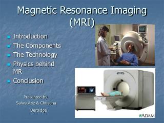
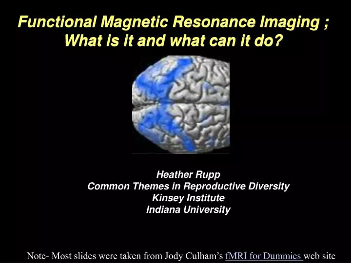
Functional Magnetic Resonance Imaging ; What is it and what can it do?. Heather Rupp Common Themes in Reproductive Diversity Kinsey Institute Indiana University. Note- Most slides were taken from Jody Culham’s fMRI for Dummies web site. New York Times September 26, 2006

E N D
Functional Magnetic Resonance Imaging ; What is it and what can it do? Heather Rupp Common Themes in Reproductive Diversity Kinsey Institute Indiana University Note- Most slides were taken from Jody Culham’s fMRI for Dummies web site
New York Times September 26, 2006 Is Hysteria Real? Brain Images Say Yes By ERIKA KINETZ Hysteria is a 4,000-year-old diagnosis that has been applied to no mean parade of witches, saints and, of course, Anna O. But over the last 50 years, the word has been spoken less and less. The disappearance of hysteria has been heralded at least since the 1960’s. What had been a Victorian catch-all splintered into many different diagnoses. Hysteria seemed to be a vanished 19th-century extravagance useful for literary analysis but surely out of place in the serious reaches of contemporary science. … New York Times September 10, 2006, Sunday The Basics; An Image of Consciousness Creates a Stir By BENEDICT CAREY (NYT) ABSTRACT - Neuroscientists were anxious as well as exuberant over the report last week that doctors in England had found clearsigns of awareness in a brain-damaged woman who was in a vegetative state. They insisted that the breathtaking finding -- that a brain thought to be all but dark flared with ... May 8, 2006 Research finds differences in lesbian brains p.m. ET May 8, 2006 WASHINGTON - Lesbians’ brains react differently to sex hormones than those of heterosexual women. An earlier study of gay men also showed their brain response was different from straight men — an even stronger difference than has now been found in lesbians…..
Today’s Goals I. What does brain imaging actually measure? MRI fMRI II. Experimental Design Basics Some Considerations III. Data Units of measurement Basic Analysis
Measuring Brain Function • Phrenology • Lesions • EEG/ERP • Electrophysiology • Need to balance considerations of spatial resolution, temporal resolution, and invasiveness.
most likely explanation: nuclear has bad connotations History of NMR • NMR = nuclear magnetic resonance • Felix Block and Edward Purcell • 1946: atomic nuclei absorb and re-emit radio frequency energy • 1952: Nobel prize in physics • nuclear: properties of nuclei of atoms • magnetic: magnetic field required • resonance: interaction between magnetic field and radio frequency Bloch Purcell NMR MRI: Why the name change?
How do you take a ‘picture’ of a brain? • Take advantage of the high (and variable) water composition of human tissue. • Hydrogen protons align with magnetic field. • Disrupt field and measure return (T1)- different brain regions vary.
1 Tesla (T) = 10,000 Gauss • Earth’s magnetic field = 0.5 Gauss • 4 Tesla = 4 x 10,000 0.5 = 80,000X Earth’s magnetic field Main field = B0 Robarts Research Institute 4T B0 x 80,000 = Source: www.spacedaily.com The Big Magnet Very strong Continuously on
Protons align with field Outside magnetic field • randomly oriented Inside magnetic field • spins tend to align parallel or anti-parallel to B0 • net magnetization (M) along B0 • spins precess with random phase • no net magnetization in transverse plane • only 0.0003% of protons/T align with field M longitudinal axis Longitudinal magnetization transverse plane M = 0 Source: Mark Cohen’s web slides Source: Robert Cox’s web slides
B0 B1 RFExcitation • Excite Radio Frequency (RF) field • transmission coil: apply magnetic field along B1 (perpendicular to B0) for ~3 ms • oscillating field at Larmor frequency • frequencies in range of radio transmissions • B1 is small: ~1/10,000 T • tips M to transverse plane – spirals down • analogies: guitar string (Noll), swing (Cox) • final angle between B0 and B1 is the flip angle Transverse magnetization Source: Robert Cox’s web slides
T1 and TR • T1 = recovery of longitudinal (B0) magnetization • used in anatomical images • ~500-1000 msec (longer with bigger B0) • TR (repetition time) = time to wait after excitation before sampling T1 Source: Mark Cohen’s web slides
T2 and TE T2 = decay of transverse magnetization TE (time to echo) = time to wait to measure T2 or T2* (after refocusing with spin echo or gradient echo) Source: Mark Cohen’s web slides
How do you take a ‘picture’ of a brain? • 1) Put subject in big magnetic field (leave him there) • 2) Transmit radio waves into subject [about 3 ms] • 3) Turn off radio wave transmitter • 4) Receive radio waves re-transmitted by subject • Manipulate re-transmission with magnetic fields during this readout interval [10-100 ms: MRI is not a snapshot] • 5) Store measured radio wave data vs. time • Now go back to 2) to get some more data • 6) Process raw data to reconstruct images • 7) Allow subject to leave scanner (this is optional) Source: Mark Cohen’s web slides
MRI vs. fMRI Functional MRI (fMRI) studies brain function. MRI studies brain anatomy. Source: Jody Culham’s fMRI for Dummies web site
History of fMRI fMRI -1990: Ogawa observes BOLD effect with T2* blood vessels became more visible as blood oxygen decreased -1991: Belliveau observes first functional images using a contrast agent -1992: Ogawa et al. and Kwong et al. publish first functional images using BOLD signal Ogawa
First Functional Images Flickering Checkerboard OFF (60 s) - ON (60 s) -OFF (60 s) - ON (60 s) - OFF (60 s) Source: Kwong et al., 1992
How do you make a ‘movie’ brain function? • Don’t look at T1 (recovery to magnetic field orientation), look at relaxation away from field T2, T2* • Relaxation differs locally and with changes in blood flow
BOLD signal Blood Oxygen Level Dependent signal • neural activity blood flow oxyhemoglobin T2* MR signal Source: fMRIB Brief Introduction to fMRI
Hemodynamic Response Function • % signal change • = (point – baseline)/baseline • usually 0.5-3% • initial dip • -more focal and potentially a better measure • -somewhat elusive so far, not everyone can find it • time to rise • signal begins to rise soon after stimulus begins • time to peak • signal peaks 4-6 sec after stimulus begins • post stimulus undershoot • signal suppressed after stimulation ends
Summary: What Does fMRI Measure? • Big magnetic field • protons (hydrogen molecules) in body become aligned to field • RF (radio frequency) coil • radio frequency pulse • knocks protons over • as protons realign with field, they emit energy that coil receives (like an antenna) • Gradient coils • make it possible to encode spatial information • MR signal differs depending on • concentration of hydrogen in an area (anatomical MRI) • amount of oxy- vs. deoxyhemoglobin in an area (fMRI)
Summary: MRI vs. fMRI MRI fMRI high resolution (1 mm) low resolution (~3 mm but can be better) one image • fMRI • Blood Oxygenation Level Dependent (BOLD) signal • indirect measure of neural activity … many images (e.g., every 2 sec for 5 mins) neural activity blood oxygen fMRI signal Source: Jody Culham’s fMRI for Dummies web site
Today’s Goals I. What does brain imaging actually measure? MRI fMRI II. Experimental Design Basics Some Considerations III. Data Units of measurement Basic Analysis
fMRI Experiment Stages: Prep • 1) Prepare subject • Consent form • Safety screening • Instructions • 2) Shimming • putting body in magnetic field makes it non-uniform • adjust 3 orthogonal weak magnets to make magnetic field as homogenous as possible • 3) Sagittals • Take images along the midline to use to plan slices Note: That’s one g, two t’s Source: Jody Culham’s fMRI for Dummies web site
fMRI Experiment Stages: Anatomicals • 4) Take anatomical (T1) images • high-resolution images (e.g., 1x1x2.5 mm) • 3D data: 3 spatial dimensions, sampled at one point in time • 64 anatomical slices takes ~5 minutes Source: Jody Culham’s fMRI for Dummies web site
first volume (2 sec to acquire) fMRI Experiment Stages: Functionals • 5) Take functional (T2*) images • images are indirectly related to neural activity • usually low resolution images (3x3x5 mm) • all slices at one time = a volume (sometimes also called an image) • sample many volumes (time points) (e.g., 1 volume every 2 seconds for 150 volumes = 300 sec = 5 minutes) • 4D data: 3 spatial, 1 temporal … Source: Jody Culham’s fMRI for Dummies web site
Cognitive subtraction originated with reaction time experiments (F. C. Donders, a Dutch physiologist). Measure the time for a process to occur by comparing two reaction times, one which has the same components as the other + the process of interest. Subtraction Logic Example: T1: Hit a button when you see a light T2: Hit a button when the light is green but not red T3: Hit the left button when the light is green and the right button when the light is red T2 – T1 = time to make discrimination between light color T3 – T2 = time to make a decision Assumption of pure insertion: You can insert a component process into a task without disrupting the other components. Widely criticized
Change only one thing between conditions! • Two paired conditions should differ by the inclusion/exclusion of a single mental process • How do we control the mental operations that subjects carry out in the scanner? • Manipulate the stimulus • works best for automatic mental processes • Manipulate the task • works best for controlled mental processes • DON’T DO BOTH AT ONCE!!! Source: Nancy Kanwisher
Add a “one back” task • subject must hit a button whenever a stimulus repeats • the repetition detection is much harder for the scrambled shapes • any activation for the intact shapes cannot be due only to attention Time Dealing with Attentional Confounds fMRI data seem highly susceptible to the amount of attention drawn to the stimulus or devoted to the task. How can you ensure that activation is not simply due to an attentional confound? Add an attentional requirement to all stimuli or tasks. • Other common confounds that reviewers love to hate: • eye movements • motor movements
Blocked Design Block Designs = trial of one type (e.g., face image) = trial of another type (e.g., place image) Assumption: Because the hemodynamic response delays and blurs the response to activation, the temporal resolution of fMRI is limited. WRONG!!!!!
Blocked vs. Event-related Source: Buckner 1998
Some Considerations Source: Doug Noll’s online tutorial
Some Considerations Average cost of performing an fMRI experiment in 1998: US$463/hour!!! Average cost of performing a thought experiment: Your Salary CONCLUSION: Unless you are Bill Gates or Michael Jordan, a thought experiment is much more efficient!
Magnet Safety The whopping strength of the magnet makes safety essential. Things fly – Even big things! Source: www.howstuffworks.com Source: http://www.simplyphysics.com/ flying_objects.html Source: Jody Culham’s fMRI for Dummies web site
Subject Safety • Anyone going near the magnet – subjects, staff and visitors – must be thoroughly screened: • Subjects must have no metal in their bodies: • pacemaker • aneurysm clips • metal implants (e.g., cochlear implants) • interuterine devices (IUDs) • some dental work (fillings okay) • Subjects must remove metal from their bodies • jewellery, watch, piercings • coins, etc. • wallet • any metal that may distort the field (e.g., underwire bra) • Subjects must be given ear plugs (acoustic noise can reach 120 dB) This subject was wearing a hair band with a ~2 mm copper clamp. Left: with hair band. Right: without. Source: Jorge Jovicich Source: Jody Culham’s fMRI for Dummies web site
Thought Experiments • What do you hope to find? • What would that tell you about the cognitive process involved? • Would it add anything to what is already known from other techniques? • Could the same question be asked more easily & cheaply with other techniques? • Would fMRI add enough to justify the immense expense and effort? • What would be the alternative outcomes (and/or null hypothesis)? • Or is there not really any plausible alternative (in which case the experiment may not be worth doing)? • If the alternative outcome occurred, would the study still be interesting? • If the alternative outcome is not interesting, is the hoped-for outcome likely enough to justify the attempt? • What would the headline be if it worked? • What are the possible confounds? • Can you control for those confounds? • Has the experiment already been done?
Today’s Goals I. What does brain imaging actually measure? MRI fMRI II. Experimental Design Basics Some Considerations III. Data Units of measurement Basic Analysis
The Data Unit VOXEL (Volumetric Pixel) Slice Thickness e.g., 6 mm In-plane resolution e.g., 192 mm / 64 = 3 mm 3 mm 6 mm SAGITTAL SLICE IN-PLANE SLICE 3 mm Number of Slices e.g., 10 Matrix Size e.g., 64 x 64 Field of View (FOV) e.g., 19.2 cm Source: Jody Culham’s fMRI for Dummies web site
Data Unit ROI Time Course fMRI Signal (% change) ~2s Condition Time Condition 1 Statistical Map superimposed on anatomical MRI image Condition 2 ... Region of interest (ROI) ~ 5 min Functional images Time Source: Jody Culham’s fMRI for Dummies web site
Averaged Over Trials Single trials Average of all trials from 2 runs
Activation is Averaged Source: Posner & Raichle, Images of Mind
Talairach & Tournoux, 1988 • squish or stretch brain into “shoe box” • extract 3D coordinate (x, y, z) for each activation focus Brain Averaging Individual brains are different shapes and sizes… How can we compare or average brains? Note: That’s TalAIRach, not TAILarach! Source: Brain Voyager course slides
What do the pretty pictures mean? Source: Jody Culham’s fMRI for Dummies web site
1. "Brain Area X is activated by Task A." • Compared to what? Activations are differences! • 2. "Baseline". • Huh?! There's a role for this, but be careful. • 3. Inferring: Because Region X responded significantly more strongly in Task A than control, but didn't respond significantly more strongly in Task B than control, it is selectively activated by Task A. • A difference in significances is not necessarily a significant difference. • 4. Imputing a specific function to a region of cortex from a difference in only two conditions. • Data always underdetermines theory, but reasonable hypotheses about function require multiple tests applied to the same region of cortex. • 5. "Gyrus X was active in my comparison of tasks B and C, and in Joe Shmo's comparison of tasks D and E, so the same area must be involved in both tasks B and D." • Gyri can be very big places; need within-subject data. Careful! Source: Nancy Kanwisher
Next Time • Meet in Room 130 Psychology • Volunteers? • http://www.indiana.edu/~imaging/index.html
Top Ten Things Sex and Brain Imaging Have in Common 10. It's not how big the region is, it's what you do with it. 9. Both involve heavy PETting. 8. It's important to select regions of interest. 7. Experts agree that timing is critical. 6. Both require correction for motion. 5. Experimentation is everything. 4. You often can't get access when you need it. 3. You always hope for multiple activations. 2. Both make a lot of noise. 1. Both are better when the assumption of pure insertion is met. Source: students in the Dartmouth McPew Summer Institute