Download
1 / 18
180 likes | 323 Views
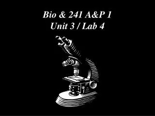
Bio & 241 A&P 1 Unit 3 / Lab 4. Histology Slides for Muscle Tissues. Slides are presented in order of magnification if different view are presented. As you view the following slides, make sure you can accomplish these goals: Can you identify the tissue observable on the slides?

E N D
Histology Slides for Muscle Tissues • Slides are presented in order of magnification if different view are presented. • As you view the following slides, make sure you can accomplish these goals: • Can you identify the tissue observable on the slides? • Can you identify the specific structures or layers indicated by the numbered arrows or brackets? • At the end of a sequence, you will find the answers to the above for each slide.
Type of Muscle Tissue? 1 3 2 1 400X
Type of Muscle Tissue? 1 2 4 400X
Type of Muscle Tissue? 6 5 5 7 400X
Type of Muscle Tissue? 9 10 100X
Type of Muscle Tissue? 9 10 11 400X
Answers to Slides 2 through 7 Slide 2: Skeletal Muscle showing both longitudinal and transverse views Slide 3: Skeletal Muscle in longitudinal view Slide 4: Cardiac Muscle in longitudinal view Slide 5: Individual smooth muscle cell Slide 6: Transverse section of the GI tract showing muscle layers Slide 7: High power view of transverse section of the GI tract showing muscle layers Numbered arrows: • Peripheral nuclei of skeletal muscle cells. Note multiple nuclei associated with each cell • A and I bands of a skeletal muscle fiber • Blood vessel in the perimysium • Perimysium between muscle fascicles • Intercalated discs • Branching fibers
Answers to Slides 2 through 7 • Centrally located nucleus • Isolated single smooth muscle cells with nucleus • Longitudinal smooth muscle layer of GI tract wall • Circular smooth muscle layer of the GI tract wall • Spindle-shaped smooth muscle cells
100X 14 13 15 12 16 14
400X 15 13 15
100X 18 17 19 20 20 13
400X 18 20 2
Answers to Slides 10 through 13 Slide 10: Muscle-Tendon intersection low power Slide 11: Muscle-Tendon intersection high power Slide 12: Neuromuscular junction low power Slide 13: Neuromuscular junction high power Numbered arrows: 12. Skeletal Muscle • Skeletal muscle cell • Epimysium surrounding muscle • Endomysium joining the tendon • Tendon • Portion of a Motor unit of a muscle • Motor nerve • Axon from a motor nerve • Neuromuscular junction with Ach vesicles
3 5 3 4 1 2 5 6
5 6 7 7
Answers to slides 15 through 13 Slide 15-16: Nerve artery vein slide with Masson stain for connective tissue Numbered Arrows: • Transverse section through a nerve • Vein • Transverse section through a muscle fascicle • Epimysium covering the muscle • Perimysium surrounding a muscle fascicle • Adipose Tissue • Endomysium surrounding muscle fibers