Hypersensitivity
Hypersensitivity. A damage to the host, mediated by preexisting immunity to self or foreign antigen. Dr. Sudheer Kher. What is hypersensitivity?. Injurious consequences in the sensitized host, following contact with specific antigen

Hypersensitivity
E N D
Presentation Transcript
Hypersensitivity A damage to the host, mediated by preexisting immunity to self or foreign antigen. Dr. Sudheer Kher Kher
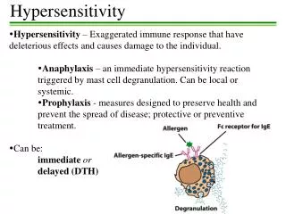
What is hypersensitivity? • Injurious consequences in the sensitized host, following contact with specific antigen • Deals with injurious aspect of heightened and exaggerated immune response leading to tissue damage, disease or even death • Concerned with what happens to the host rather than what happens to the antigen. Kher
Musts for Hypersensitivity • Contact with allergen • Sensitizing/priming dose • Induction of AMI/CMI • Shocking dose Kher
Classification:Hypersensitivity reactions • Immediate hypersensitivity • Anaphylaxis • Atopy • Antibody mediated cell damage • Arthus phenomenon • Serum sickness • Delayed hypersensitivity • Infection (Tuberculin) type • Contact dermatitis type Kher
Type I (Anaphylactic) Reactions • Occur within minutes of exposure to antigen • Antigens combine with IgE antibodies bound to mast cells and basophils, causing them to undergo degranulation and release several mediators: • Histamine: Dilates and increases permeability of blood vessels (swelling and redness), increases mucus secretion (runny nose), smooth muscle contraction (bronchi). • Prostaglandins: Contraction of smooth muscle of respiratory system and increased mucus secretion. • Leukotrienes: Bronchial spasms. • Anaphylactic shock: Massive drop in blood pressure. Can be fatal in minutes.
Type I Reactions ( IgE Mediated) • Anaphylaxis – • Classical immediate reaction • Sensitization • Most effective when Ag introduced parenterally • May occur by any route exposure to Ag • Minute quantities are enough • Interval of 2-3 wks needed between sensitizing & shocking dose • Once sensitized it remains so for long time • Shocking dose most effective by IV route then IP, then SC then ID • The shocking Ag must be same or similar to Sensitizing Ag Kher
B cell IL13 TH2 Newly synthesized mediators Sensitization against allergens and type-I hypersensitivity Histamine, tryptase, kininegenase, ECFA Leukotriene-B4, C4, D4, prostaglandin D, PAF Kher
Type I Reactions • Humans – • Itching of scalp & tongue, flushing of skin, difficulty in breathing, nausea, vomiting, diarrhea, acute hypotension, loss of consciousness, death (rare) • Causes • Serum therapy, antibiotics, insect stings • Treatment • Adrenalin 0.5 ml (1 in 1000 solution) SC/IM repeated up to 2 ml in 15 min Kher
Cutaneous anaphylaxis • If small shocking dose is given ID to sensitized host, there is a local weal & flare reaction (local anaphylaxis). • Used for • Testing for hypersensitivity • Identification of allergens for atopy • Precaution – Keep adrenalin injection ready to combat severe fatal reaction. Kher
Mechanism of anaphylaxis • Mediators of anaphylaxis – • Primary mediators • Preformed contents of Mast cells & Basophils • Histamine, serotonin, eosinophils chemotactic factor of anaphylaxis (ECF-A), Neutrophil chemotactic factor (NCF), Heparin & various proteolytic enzymes • Secondary mediators – • Newly formed after stimulation by Mast cells, Basophils & other leucocytes • Slow reacting substance of anaphylaxix (SRS-A), Prostaglandins & Platelet activating factors (PAF) Kher
Primary Mediators of Anaphylaxis • Histamine – • Most important vasoactive amine of Human anaphylaxis, formed from histidine found in granules. Released into skin, causes burning & itching. Causes vasodilatation & hyperemia by an axon reflex (Flare) and edema by increasing capillary permeability (Weal). Induces smooth muscle contraction of diverse tissues & organs. Kher
Primary Mediators of Anaphylaxis • Serotonin (5-HT) – • Base derived by decarbolxylation of Tryptophan. • Found in intestinal mucosa, brain & platelets. • Causes smooth muscle contraction, ↑ Vascular permeability & vasoconstriction. • Important in rats & mice. • Role in human not clear. Kher
Primary Mediators of Anaphylaxis • Chemotactic factors – • ECF-A released from mast cell granules are strongly chemotactic for eosinophils. Accounts for high eosinophil counts in many hypersensitivity reactions. • NCF – Attracts neutrophils • Heparin – Acidic mucopolysaccharide. Contributes to anaphylaxis in dogs but apparently not in man. • Enzymatic mediatores such as proteases & hydrolases are also released from the mast cell granules. Kher
Secondary mediators of anaphylaxis • Prostaglandins & leukotrienes – • Derived from Arachidonic acid formed from the disruption of mast cell membrane other leucocytes • Lipoxygenase pathway - Leukotrienes • Cycloxygenase pathway - Prostaglandins • One of the family of Leukotrienes is SRS-A (slow reacting substance of anaphylaxis) • Prostaglandins are bronchoconstrictors/broncodilators, affect secretions of mucus glands, platelet adhesion, permeability, dilatation of capillaries & pain threshold. Kher
Secondary mediators of anaphylaxis • Platelet activating factor – PAF • Low mol wt lipid released from basophils • Causes aggregation of platelets and release of their vasoactive amines • Other mediators – • Anaphylatoxin – Released by complement activation • Bradykinin & Other kinins formed from plasma kininigens Kher
Type-I hypersensitivity The common allergy Kher
Anaphylactoid reaction • Intravenous injection of peptone, trypsin & certain other substances causes clinical reaction like anaphylaxis. • Resemblance due to participation of same chemical mediators. • Difference – Anaphylactoid shock has no immunological basis. It is nonspecific reaction involving activation of complement & release of anaphylatoxin. Kher
Type II (Cytotoxic) Reactions • Involve activation of complement by IgG or IgM binding to an antigenic cell. • Antigenic cell is lysed. • Transfusion reactions: • ABO Blood group system: Type O is universal donor. Incompatible donor cells are lysed as they enter bloodstream. • Rh Blood Group System: 85% of population is Rh positive. Those who are Rh negative can be sensitized to destroy Rh positive blood cells. • Hemolytic disease of newborn: Fetal cells are destroyed by maternal anti-Rh antibodies that cross the placenta.
Type II Reactions : Cytolytic, Cytotoxic & Cell Stimulatory • Involve reaction between IgG (rarely IgM) & Ag determinant on the surface of cells. • Leads to cytolytic or cytotoxic effect. • Autoimmune anemias • Hemolytic disease of the new born • Drug induced hemolytic anemias • Drug induced thrombocytopenic purpura • Drug induced agranulocytosis • Rarely normal cell function may be disrupted • Agonist effect -Cell stimulatory effect seen (LATS in Graves’ disease). • Antagonist effect – Myasthenia gravis Kher
Type III (Immune Complex) Reactions • Involve reactions against soluble antigens circulating in serum. • Usually involve IgA antibodies. • Antibody-Antigen immune complexes are deposited in organs, activate complement, and cause inflammatory damage. • Glomerulonephritis: Inflammatory kidney damage. • Occurs when slightly high antigen-antibody ratio is present.
Type III Reaction: Immune Complex Disease • Arthus Reaction – Localized manifestation of generalized hypersensitivity • Ag+Ab precipitates cause C activation and release of inflammatory molecules. Leads to ↑ vascular permeability & neutrophil infiltrate. Leucocyte-platelet thrombi formed which reduce blood supply leading to necrosis. • Clinical example – Farmer’s lung & other hypersensitivity pneumonitis following inhaled Ag like Actinomycetes. Kher
Arthus reaction Arthus reaction Type-III Weal & flare reaction Type-I Kher
Type III Reaction: Immune Complex Disease • Serum Sickness – Systemic form of Type III reaction. • Takes place following serum therapy • e.g. ADS, ATS, AGGS, Hyperimmune globulin, Anti Snake venum. • Clinically Fever, lymphadenopathy, splenomegaly, arthritis, glomerulonephritis, endocarditis, vasculitis, urticarial rashes, abdominal pain, nausea, vomiting. • Pathogenesis – Formation of immune complexes, its deposition on the endothelial lining of BVs all over the body, leads to inflammation. Kher
Serum Sickness (contd) • Plasma concentration of C falls due to massive activation and fixation to Ag+Ab complexes. • Disease self limited. • Single dose of Antiserum can serve both as sensitizing & shocking dose. • Can also be seen after administration of penicillin or other antibiotics. • Immune complexes occur in many bacterial, viral, parasitic infections e.g. poststreptococcal glomerulonephritis, Hepatitis B & Malaria. Also seen in disseminated malignancies & autoimmunity. Kher
Serum sickness Kher
Type IV (Cell-Mediated) Reactions • Involve reactions by TD memory cells. • First contact sensitizes person. • Subsequent contacts elicit a reaction. • Reactions are delayed by one or more days (delayed type hypersensitivity). • Delay is due to migration of macrophages and T cells to site of foreign antigens. • Reactions are frequently displayed on the skin: itching, redness, swelling, pain. • Tuberculosis skin test • Poison ivy • Metals • Latex in gloves and condoms (3% of health care workers) • Anaphylactic shock may occur.
Type IV Reactions: Delayed Hypersensitivity • One aspect of CMI • Provoked by specific Ag, involves lymphocytes & Macrophages. • Not induced by circulating Ab but by sensitized lymphocytes. • Sensitized lymphocytes release lymphokines which have biological effects on leucocytes, macrophages & tissue cells. • Transfer possible thru’ lymphocytes / transfer factor. Kher
Type IV Reactions: Delayed Hypersensitivity • Two types – • Tuberculin (Infection) type • Contact dermatitis type • Tuberculin type – • ID inoculation of PPD in sensitized indivisual leads to induration & inflammation in 48-72 hrs. This is not same as skin test done for Type I hypersensitivity. • Used for diagnosis / exclusion of diagnosis of many bacterial / fungal / parasitic / viral and autoimmune diseases. Kher
Contact dermatitis • Ag possibly enters thru’ sebaceous glands • Lesions vary from macules & papules to vesicles which subsequently breakdown leaving weeping surface typical of acute eczematous dermatitis. • Detected by patch test Kher
Allergic Contact Dermatitis Response to Poison Ivy Hapten Kher
characteristic Type-I Type-II Type-III Type-IV antibody IgE IgG, IgM IgG, IgM none exogenous cell surface soluble intracellular antigen response time 15-30 min. Min.-hrs 3-8 hours 48-72 hours or longer Erythema & edema Erythema & induration Lysis & necrosis appearance Weal & flare baso- and eosinophils Ab and complement Monocytes & lymphocytes histology PMN and complement antibody T-cells antibody transfer with antibody examples hay fever, asthma pemphigus, Goodpasture farmers’ lung, SLE TB test, poison ivy, granuloma Comparison of hypersensitivity reactions Kher