Download
1 / 39
560 likes | 877 Views
Dr. ÁgostonSzél. Clinical anatomy of the male pelvis and pelvic floor. Semmelweis University Deaprtment of Anatomy , Histology and Embryology 2019. The pelvis as a whole. The pelvis. greater pelvis = pelvis major, iliac fossa (part of the abdominal cavity, false pelvis).

E N D
Dr. ÁgostonSzél Clinicalanatomy of themalepelvis and pelvicfloor Semmelweis University Deaprtment of Anatomy, Histology and Embryology 2019
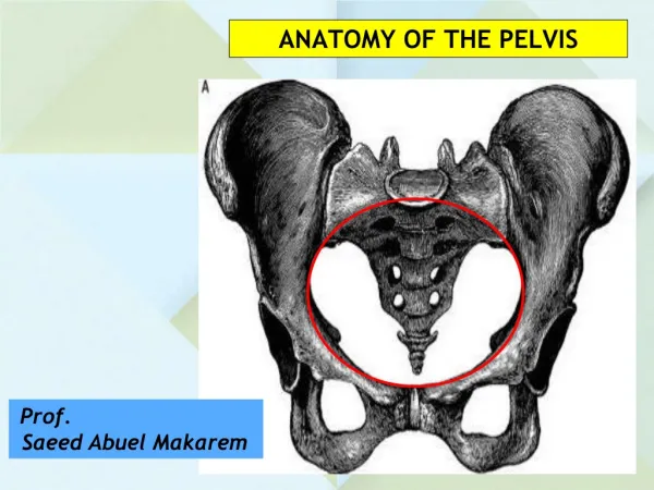
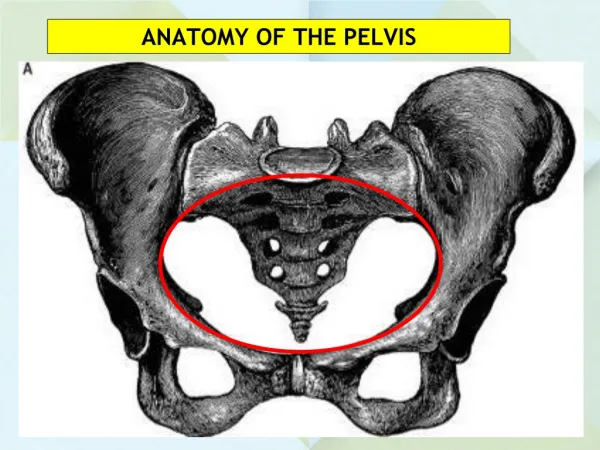
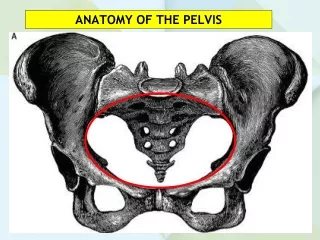
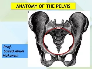
The pelvis greater pelvis = pelvis major, iliac fossa (part of the abdominal cavity, false pelvis) promontory borderline = linea terminalis pubic symphysis lesser pelvis = pelvis minor, true pelvis
Bonyconstituents of thepelvis promontory linea terminalis ischial spine pubic tubercle sacrococcygeal joint pubic torus symphysis lower edge of symphysis ischial tuberosity
posterior sacroiliac ligament interosseal sacroiliac ligaments iliac bone sacrum sacroiliac joint joint cavity cartilagineous discs sacrotuberous ligament greater sciatic foramen sacrospinous ligament anterior sacroiliac ligament lesser sciatic foramen ant. and post. symphyseal ligaments cartilagineous disks pubic bone symphysis fibrocartilagineous discs
Bones and ligaments of pelvis intervertebral foramen L5 lumbosacral joint sacrum iliac crest iliac fossa ant. sup. iliac spine sacroiliac joint INGUINAL LIGAMENT 1. subinguinal hiatus ant. inf. iliac spine 2. greater sciatic foramen arcuate line SACROSPINOUS LIGAMENT linea terminalis pecten of pubic bone coccygeal bone 3. lesser sciatic foramen pubic tubercle SACROTUBEROUS LIGAMENT 5. inferior pelvic aperture symphyseal surface 4. obturator foramen
Diameters of pelvis sup. pelvic aperture amplitude (wide part) of pelvis (d-h) true anatomical conjugate (a-b) angustia (narrow part) of pelvis (e-g) inf. pelvic aperture true obstetric conjugate (a-c) axis of pelvis (k-k) internal diagonal conjugate (a-e)
Parts of lesserpelvis (pelviccanal, truepelvis, birthcanal) amplitude (wide part) of pelvis pelvic inlet (sup. pelvic aperture) angustia (narrow part) of pelvis axis of pelvis linea terminalis interior of pelvis pelvic outlet (inf. pelvic aperture)
11 cm 12 cm 13 cm oblique diameter transverse diameter median diameter Important diameters of pelvic inlet interspinal distance (25-26 cm) intercristal distance (28-29 cm)
Variations of theshape of pelvicinlet Classification of Caldwell & Moloy
Sexualdifferences in theshape of pelvis female male inlet outlet cavity of pelvis pubic arch
Superiorpelvicaperture heart-shaped, oval, ellipsoid Transverse diameter
Amplitude of pelvis mediane diameter circular transverse diameter
Inferiorpelvicaperture mediane diameter (conjugate) rhomboid-shaped
Openings and muscularcomponents of pelvis intervertebral foramen iliacus psoas maj. & min. suprapiriform hiatus 2. greater sciatic foramen piriformis 1. subinguinal hiatus infrapiriform hiatus muscular and nervous, lacunae, vascular lacuna, lymphatic lacuna ischiococcygeus 4. obturator foramen 3. lesser sciatic foramen obturator canal int. obturator tendinous arch (levator ani removed) 5. inferior pelvic aperture gluteus max.
Pelvisasseenfromabove urogenital & anal hiatus obturator canal pubococcygeus iliococcygeus tendinous arch rectococcygeus ischiococcygeus greater sciatic foramen sacrotuberous lig. ant. sacroccocygeus
Parts of levator ani muscle urogenital hiatus sphincter of vagina inferior ramus of pubic bone puborectalis centrum tendineum pubococcygeus ischiadic tubercle sacrotuberous ligament iliococcygeus
Levator ani muscle insertion of levator ani on the pelvic wall greater sciatic foramen sacro-spinous lig. obturator membrane sacro-tuberous lig. lesser sciatic foramen
Maintenance of theintraabdominalpressure thoracic diaphragm abdominal wall pelvic diaphragm
Maintenance of theintraabdominalpressure thoracic diaphragm abdominal wall pelvic diaphragm
Pelvicfloorasseenfrombelow urethra vagina deeptransverseperineal A superficialtransverseperineal obturator int. B obturator fascia pubococcygeus* puborectalis iliococcygeus ischiococcygeus external anal sphincter centrum tendineum external anal sphincter anococcygeal lig. pubovaginalis* levator of prostate levator ani*
Parts of levator ani muscle rectum symphysis perineal flexure puboprostatic lig. puborectalis levator of prostate
Parts of levator ani muscle urogenital hiatus levator of prostate pubovaginalis (sphincter of vagina) inf.ramus of pubic bone puborectalis centrum tendineum pubococcygeus ischiadic tubercle iliococcygeus sacrotuberous lig.
Pararectalspace (paraproctium) and fasciae rectal fascia proper slows down the propagation of metastatic prostate cancer* (visceral pelvic fascia) presacral fascia rectoprostatic fascia* (Denonvilliers) (parietal pelvic fascia) sup. rectosacral space** rectoprostatic space (Denonvilliers) rectosacral fascia (Waldeyer) inf. rectosacral space inf.** space without blood and lymph vessels („holy plane”)** rectovaginal fascia* (Denonvilliers)
peritoneum A B pelvic fascia (visceral layer) endopelvicfascia (parietallayer) frontal section at the level of urinary tract (A) frontal section at the level of rectum (B) anterior continuation of ischiorectal fossa sagittal section ischiorectal fossa
Sagittalsection of themalepelvis rectovesical pouch sigmoid colon presacralfascia ileum peritoneum rectalfasciaproper rectum peritoneal cavity rectosacralfascia (Waldeyer) bladder seminal vesicle prevesical space (Retzius)* ejaculatory duct puboprostatic lig. rectosacralspace rectoprostatic space/fascia (Denonvilliers)* prostate anococcygeal lig. urogenital diaphragm ext anal sphincter int. anal sphincter anus anal canal centrum perineum prostatic part of urethra *pre- and infra-peritoneal space *remnant of recto-vesical pouch (septum) spf. abdominal fascia (Scarpa) scrotum
obturatorinternus Schematicview of pelvicfloor and urogenitaldiaphragm (frontalsection) pelvic diaphragm (levator ani) visceral pelvic fascia sup. fascia of pelvic floor endopelvicfascia (parietalpelvicfascia) inf. fascia of pelvic floor obturatorfascia sup. fascia of urogenitaldiaphragm inf. fascia of urogenital diaphragm (perineal fascia) parietalisfasciapelvis urogenital diaphragm (deep transverse perineal muscle)
Schematicview of pelvicfloor and urogenitaldiaphragm (saggittalsection) A B pelvic floor (levator ani) urogenital diaphragm (deep transv. perineal muscle.)
Pelvic and urogenitaldiaphragm vulva deeptransv. perinealmuscle bulbocavernosus inf. ramus of pubic bone centrum tendineum ischiocavernosus spf. transv. perinealmuscle ischiadic tubercle obturator internus m. levator ani ext. anal sphincter anococcygeal lig.
symphysis subarcuate hiatus centrum tendineum arcuate ligament of pubis urethra inferior ramus ofpubic bone ischiadic tubercle urogenital diaphragm inferiorrectalartery external anal sphincter anococcygeal ligament levator ani coccygeal bone inferiorrectalnerve sacrotuberous ligament gluteus maximus
corpus of clitoris glans of clitoris crus of clitoris urethralorifice bulb of vestibule ischiocavernosus bulbospongiosus hymen inferior fascia of urogenital diaphragm (perineal membrane) greatervestibulargland superficial transverse perineal muscle centrum tendineum gluteus maximus external anal sphincter anus levator ani anococcygeal ligament
dorsalpenilevein corpus cavernosum corpus spongiosum ischiocavernosus crus of penis bulbospongiosus centrum tendineum urogenital diaphragm external anal sphincter bulb of penis gluteus maximus superficial transverse perineal muscle levator ani
pelvic fascia endopelvic fascia levator ani obturator internus prostate pelvis (infraperitoneum) prostatic part of urethrae urogenital diaphragm „ischiorectal fossa” superior fascia of urogenital diaphragm dorsalpenilenerve deepperinealspace. urogen. diaphragm, urethralsphincter, bulbourethralgland, vessels, nerves membraneous part of urethra deeppenileartery inferior fascia of urogenital diaphragm (perineal membrane) artery of bulb of penis crus of penis ischiocavernous bulbourethralgland spongy part of urethra bulb of penis bulbospongiosus superficialperinealspacescrotalnerves spf. perineal fascia (Colles) deep femoral fascia innersurface of thigh
levator ani vagina obturator fascia pelvic fascia paracolpium pelvis deep perineal space, urogenitaldiaphragm obturator internus obturator membrane „ischiorectal fossa” superior fascia of urogenital diaphragm dorsalnerve of clitoris deepartery of clitoris internalpudendalartery crus of clitoris artery of bulb inferior fascia of urogenital diaphragm (perineal membrane) ischiocavernosus superficialprinealspacelabialnerves bulb of vestibule bulbospongiosus deep femoral fascia hymen greatervestibulargland innersurface of thigh labiummajus labiumminus superficial perineal fascia (Colles)
peritoneal cavity fascia perinei spf. (Colles) Spaces of thepelvis in sketch obturatorinternus pelvic diaphragm (levator ani) peritoneum 1. pelvis infraperitoneal space 2. pudendal canal (Alcock) 3. „ischiorectal fossa” deep perineal space 4. urogenital diaphragm (deep transv. perineal) deepperinealpouch superficial perineal space ischiocavernosus bulbospongiosus
Internaliliacartery common iliac internal iliac uterine iliolumbal umbilical (sup. vesical) superior gluteal lateral sacral external iliac inferior gluteal internal pudendal obturator middle rectal inf. vesical
Internalpudendalartery internal pudendal (greater sciatic foramen) common iliac lateral sacral laterales internal iliac (pelvic sacral foramens) externaliliac femoral internal pudendal (lesser sciatic foramen) external pudendal dorsal penile (subarcuate hiatus) inf. rectal deep penile (uro-genital diaphragm) a. of bulb of penis perineal post. scrotal ant. scrotal
Bibliography • Szentágothai J, Réthelyi M: Funkcionális anatómia. Medicina, 1985. • Lippert H: Lehrbuch Anatomie, Urban & Fischer, München, 2000. • Szél Á: Klinikai anatómia, Semmelweis, Budapest, 1999. • Snell RS: Clinical Anatomy, Little, Brown & Co, Boston, 1995. • Moore KL, Dalley AF: Clinically Oriented Anatomy, Lippincott, 1999. • Renz-Polster H, Krautzig S, Braun J: Innere Medizin, Urban & Fischer, München, 2004. • Putz R, Pabst R: Sobotta. Alliter, Budapest, 2004. • Patel R: Applied Peritoneal Anatomy, www.myESRorg. • Tirkes T et al: Peritoneal Anatomy. Gastrointestinal Imaging, https://doi.org/10.1148/rg.322115032.