Download
1 / 80
1.06k likes | 1.57k Views
Anatomy of the Female Pelvis. By DR. Areefa Albahri Assistance Prof. of MCH Islamic University of Gaza. The primary function of the pelvic girdle is to allow movement of the body, especially walking and running.

E N D
DR. Areefa Albahri Anatomy of the Female Pelvis By DR. Areefa Albahri Assistance Prof. of MCH Islamic University of Gaza
DR. Areefa Albahri • The primary function of the pelvic girdle is to allow movement of the body, especially walking and running. • It permit the person to sit and kneel. The women pelvis is adapted for child bearing, because of its increased width and rounded brim women are less speedy than men. The pelvis afford protection to the pelvis organ
The female external reproductive system • The female reproductive system consists of the external genitalia, known collectively as the vulva and the internal reproductive organs: the vagina, the uterus, two uterine tubes and two ovaries. In the non-pregnant state, the internal reproductive organs are situated within the true pelvis. • The vulva • The vulva includes the • The mons pubis is rounded pad of fat lying over the symphysis pubis. It is covered with pubic hair from the time of puberty.
The female external reproductive system • The labia majora(‘greater lips’) are two folds of fat and areolar tissue covered with skin and pubic hair on the outer surface and have pink smooth inner surface. • The labia minora(‘lesser lips’) are two thin folds of skin lying between the labia majora. Anteriorly they divide to enclose the clitoris; the frenum is formed by the two medial parts; posteriorly they fuse, forming the fourchette.
The clitoris is a small rudimentary sexual organ corresponding to the male penis; the visible knob-like portion is located near the anterior junction of the labia minora, above the opening of the urethra and vagina. The prepuce a retractable piece of skin surrounds and protects the clitoris. Unlike the penis, the clitoris does not contain the distal portion of the urethra and functions solely to induce the orgasm of sexual intercourse.
External genitalia mons pubis Labium majus Labium minus Urethral orifice Vaginal orifice Vaginal vestibule Perineal body Anus
The vestibule is the area enclosed by the labia minora in which the openings of the urethra and the vagina are situated. • The urethral orifice lies 2.5 cm posterior to the clitoris and immediately in front of the vaginal orifice. • The vaginal orifice,Theorifice is partially closed by the hymen, a thin membrane that tears during sexual intercourse or during the birth of the first child. • Bartholin's glands are two small glands They secrete mucus, which lubricates the vaginal opening.
Blood supply • The blood supply comes from the internal and the external pudendal arteries. The blood drains through corresponding veins. • Lymphatic drainage • Lymphatic drainage is mainly via the inguinal glands. • Nerve supply • The nerve supply is derived from branches of the pudendal nerve. The vaginal nerves supply the erectile tissue of the vestibular bulbs and clitoris and their parasympathetic fibres have a vasodilator effect.
The perineum • The perineum to the outer of the pelvis and is some what lozenge- shape : anteriorly it is bounded by the pubic arch , posteriorly by the coccyx, and laterally by the ischiopubic rami, ischialtuberosities and sacrotuberous ligament • The perineum divided into two triangular part: • 1. the urogenital triangle • 2. the anal triangle
The pelvic floor • The pelvic floor or pelvic diaphragm is composed of muscle fibers of the levatorani,(paired levatorani muscle LAM) the coccygeus muscle, and associated connective tissue which span the area underneath the pelvis. The pelvic diaphragm is a muscular partition formed by the levatoresani and coccygei, with which may be included the parietal pelvic fascia on their upper and lower aspects. The pelvic floor separates the pelvic cavity above from the perineal region (including perineum) below.
Function of levator muscles • 1.maintain constant tone, except during voiding defecation • 2. have the ability to contract quickly at the time of acute stress such as cough and sneeze • 3.distended considerably during delivery
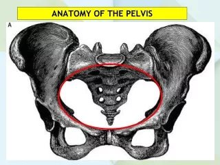
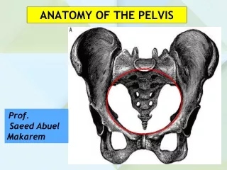
The pelvis • The pelvic girdle, a basin shaped cavity, and consist of two innominate bone (hip bones), the sacrum and the coccyx. It is also a bony ring between the movable vertebrae of the vertebral column which it supports, and the lower limbs that it rests on. It contains and protects the bladder, rectum and internal reproductive organs. Some women experience pelvic girdle pain in pregnancy and need referral to an obstetric physiotherapist
Pelvis False pelvis (pelvis major) True pelvis (pelvis minor)
Innominate bone • Each innominate bone or( hip bone) is mad up of three bones have fused together : the ilium, ischium, and pubis. It is fixed bone.
HIP BONE Ileum Pubis Ischium
The ilium • has an upper & lower part . The smaller lower part form part of acetabulum and the upper part is the large flared out part. When the hands is placed on the hip it rests on the iliac crest which known as anterior superior iliac spine.
The ischium • is the inferoposterior part of the innomiate bone and consist of a body and ramus. Above it form part of acetabulum. It has a large prominence known as the ischial tuberosity on which the body rests when sitting. Behind and a little above the tuberosity is an inward projection, the ischial spine. In labour, the station of the fetal head is estimated in relation to the ischial spines.
The pubis • forms the anterior part. It has a body and two oar-like (blade) projections, the superior ramus and the inferior ramus. The two pubic bones meet at the symphysis pubis and the two inferior rami form the pubic arch, merging into a similar ramus on the ischium. The space enclosed by the body of the pubic bone, the rami and the ischium is called the obturator foramen. The innominate bone contains a deep cup to receive the head of the femur termed the acetabulum, which is composed of the three fused bones in the following proportions: two-fifths ilium, two-fifths ischium and one-fifth pubis
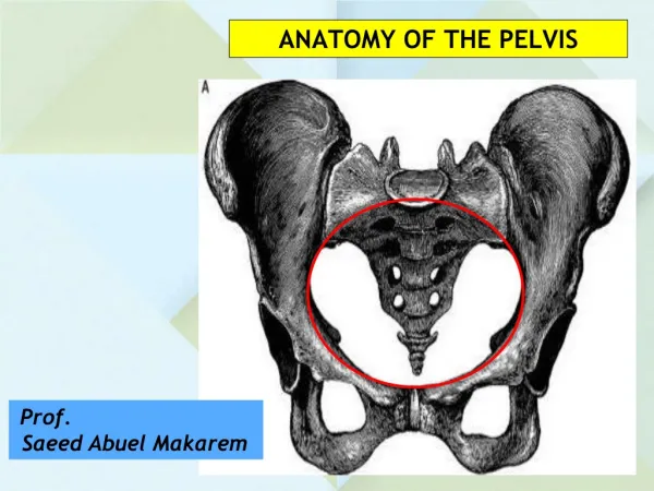
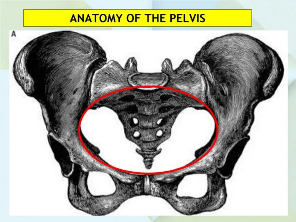
The sacrum • The sacrum is a wedge-shaped bone consisting of five fused vertebrae. The upper border of the first sacral vertebra, which juts forward, is known as the sacral promontory. The anterior surface of the sacrum is concave and is referred to as the hollow of the sacrum. nerves from the caudaequina emerge to supply the pelvic organs. The posterior surface is roughened to receive attachments of muscles.
Sacral promontory 2 Innominate bone sacrum coccyx Ischail spine Symphsis pubis
The coccyx • The coccyx is a vestigial tail. It consists of four fused vertebrae, forming a small triangular bone, which articulates with the fifth sacral segment.
Pelvic joints • Pelvic joints • There are four pelvic joints: • 1. one symphysis pubis • 2. two sacroiliac joints • 3 one sacrococcygeal joint. • The symphysis pubis is the midline cartilaginous joint uniting the rami of the left and right pubic bones. • The sacroiliac joints are strong, weight-bearing synovial joints. They join the sacrum to the ilium and as a result connect the spine to the pelvis. The joints allow a limited backward and forward movement of the tip and promontory of the sacrum, sometimes known as ‘nodding’ of the sacrum. • The sacrococcygeal joint is formed where the base of the coccyx articulates with the tip of the sacrum. It permits the coccyx to be deflected backwards during the birth of the fetal head.
Pelvic ligaments • The ligaments connecting the bones of the pelvis with each other can be divided into four groups: • Sacroiliac ligament • those connecting the sacrum and ilium – the sacroiliac ligaments • Sacrospinous ligament • those passing between the sacrum and ischium – • Sacrococcygeal ligaments • those uniting the sacrum and coccyx • interpubic ligaments • those between the two pubic bones The ligaments that are important to midwifery practice are the sacrotuberous and the sacrospinous ligaments as they form the posterior wall of the pelvic outlet
Classification of pelvis Divided into: 1) False pelvis (pelvis major; greater pelvis) • Part of abdominal cavity 2) True pelvis (pelvis minor;lesser pelvis ) • Is the true pelvic cavity • Bony canal through which fetus must pass during labor • It divided into a brim, a cavity, and outlet False pelvis True pelvis
The pelvic brim • The pelvic brim called pelvic inlet :Pelvic inlet ( = pelvic brim) • The brim is rounded except where the sacral promontory • projects into it. • The midwife need to know the fixed point on the pelvic brim that are known as the land marks
1 - Sacral Promotory 2 - Sacral ala (wing) 3 - Sacral iliac joint 4 - Illiopectineal line- 5- Illiopectineal eminence 6- Superior pubic ramus 7- Body of pubic bone 8- Symphysis pubis
The pelvic cavity • The cavity extended from the brim superior to the outlet inferiorly.
The pelvic outlet • The anatomical outlet is formed by the lower borders of each of the bones togehter with the sacrotuberous ligament. It include the narrow pelvic strait which the fetus must pass.
Inlet cavity Anterior posterior 11 cm Transfer 13 cm obliqu12 cm
The Cavity..!!! • Round cavity of greatest dimensions. • Anteroposterior diameter • Oblique diameter • Transverse diameter 12 cm
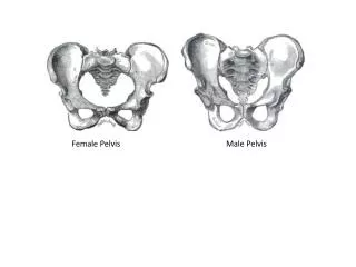
FEMALE MALE • Bones are lighter, thinner • False pelvis is shallow • Pelvic cavity is wide & shallow • Pelvic inlet round/oval • Pelvic outlet comparatively large • Subpubic angle large • Coccyx more flexible, straighter • Ischialtuberosities more everted • Bones heavier, thicker • False pelvis is deep • Pelvic cavity is narrow & deep • Pelvic inlet heart-shaped + smaller • Pelvic outlet comparatively small • Subpubic angle more acute • Coccyx less flexible, more curved • Ischialtuberosities longer, face more medially
variation of pelvic shape • Female pelvis shapes may be subdivided as follows • 1. Normal and its variants -Gynaecoid – most common type , suited for delivery - Android – the male type of pelvis - Platypelloid– flat pelvis; short AP diameter & wide transverse diameter - Anthropoid– resembling that of anthropoid ape, AP diameter is greater than the transverse 2. Symmetrically contracted pelvis - That of a small women but with a symmetrical shape
3. Rachitic pelvis - This deformity is caused by rickets (due to Vit D deficiency) - Sacrum is rotated so that the sacral promontory projects forward and coccyx tips backward - AP diameter of inlet is therefore narrowed but the outlet is increased 4. Asymmetrical pelvis - Asymmetry pelvis can be due to variety of causes such as scoliosis, poliomyelitis, pelvic fracture, congenital abnormality due to thalidomide etc Rachitic pelvis Asymmetrical pelvis
Gynaecoid pelvis • Is a typical female pelvis. Ideal for vaginal delivery • Found in 80 % of Asian women; 50-70 % white women • Rounded or slightly oval inlet • Straight pelvic sidewalls with roomy pelvic cavity • Good sacral curve • Subpubic arch is wide 90 degree
Android pelvis • Present in most male and also in few females • Heart shaped (or triangular) pelvic inlet - due to prominent sacrum • The problem in delivery head occiptoposterior most common • Narrow sub-pubic angle less than 90
Anthropoid pelvis • Present in some males and females • 15% in Asian women; 15-30% in white women • Pelvic inlet is long oval • AP diameter > transverse diameter • Long & narrow sacrum • Women with this type tend to be tall. • Less labor complications