pelvic anatomy
Learning Topics. Bony PelvisRoutes from abdomen to pelvisLayers and compartmentsFemale reproductive anatomyMale reproductive anatomyNeurovascular supplyMEQ practice examplesExtra


pelvic anatomy
E N D
Presentation Transcript
1. Pelvic Anatomy Dan Smith
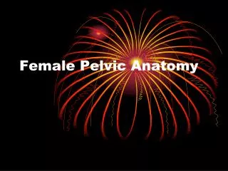
3. If we look in detail at the hip bones we find that they are made from 3 separate bones that are fused together, these bones being the ilium��..the pubic����.and the ischium. The fusing of the three bones is only complete in late puberty, or even early adulthood. At the point of fusion of the three bones we find an indented region of the hip bone known as the acetabulum. The acetabulum forms the socket part of the hip joint.
The large fan shaped bone that is located superiorly is known as the ilium. The top of the iliac bone is known as the iliac crest, and it is this piece of bone that you can feel on the lateral sides of your pelvis, near to your waist. Try to feel this piece of bone now.
The anterior part of the hip bone is formed by the pubic bone, shown on the diagram in pink. The pubic bone is found in the region we often refer to as the pubic region. The bone can be felt posterior to the area normally covered in pubic hair. If you are in an appropriate place try to feel the outline of this bone now.
The final bone contributing to the hip bone is the ischium, shown on the diagram in blue. The ischium is located relatively posteriorly and inferiorly. The lower part of the ischium is known as the ischial tuberosity, and it is through this piece of bone that weight is transferred whilst sitting.
If we look in detail at the hip bones we find that they are made from 3 separate bones that are fused together, these bones being the ilium��..the pubic����.and the ischium. The fusing of the three bones is only complete in late puberty, or even early adulthood. At the point of fusion of the three bones we find an indented region of the hip bone known as the acetabulum. The acetabulum forms the socket part of the hip joint.
The large fan shaped bone that is located superiorly is known as the ilium. The top of the iliac bone is known as the iliac crest, and it is this piece of bone that you can feel on the lateral sides of your pelvis, near to your waist. Try to feel this piece of bone now.
The anterior part of the hip bone is formed by the pubic bone, shown on the diagram in pink. The pubic bone is found in the region we often refer to as the pubic region. The bone can be felt posterior to the area normally covered in pubic hair. If you are in an appropriate place try to feel the outline of this bone now.
The final bone contributing to the hip bone is the ischium, shown on the diagram in blue. The ischium is located relatively posteriorly and inferiorly. The lower part of the ischium is known as the ischial tuberosity, and it is through this piece of bone that weight is transferred whilst sitting.
4. Pelvis
5. Pelvis
6. Pelvic Foramen Obturator Canal:
Obturator nerve and vessels.
Greater Sciatic Foramen:
Above piriformis: - superior gluteal nerves and vessels
Below piriformis: - inferior gluteal nerve and vessels
- sciatic nerve
- pudendal nerve and vessels
- nerve to obturator internus, post. femoral cutaneous nerves, nerve to quadratous femoris
Lesser Sciatic Foramen:
Pudendal nerve and vessels enter perineum
Tendon of obturator internus muscle
9. Compartments of the urogenital traingle
10. CONTENTS OF MALE SUPERFICIAL PERINEAL POUCH
11. CONTENTS OF FEMALE SUPERFICIAL PERINEAL POUCH
12. Perineal Membrane
14. Deep Perineal Pouch
15. Pelvic diaphragm = levator ani and coccygeus muscles.Pelvic floor= pelvic diaphragm, perineal membrane and muscles in the deep perineal pouch
17. Compartments of the urogenital traingle
18. Female reproductive anatomy
19. Female reproductive anatomy
20. Uterine Support Uterine support thought to be by:
Ligaments: - from the uterus to the pelvic walls
Pubocervical
Transverse cervical (cardinal ligament)
Uterosacral
Perineal membrane
Pelvic floor (especially levator ani)
Perineal body
21. Ligaments Broad Ligament:
Double fold of peritoneum extending laterally from the uterus towards the pelvic side wall and encloses the uterine tube.
Between the fold the uterine and ovarian arteries anastomose
Ovarian Ligament:
Forms a ridge on the posterior leaf of the broad ligament. It is developmentally part of the gubernaculum and in continuity with the round ligament.
Round ligament:
Curves anteriorly to pass through the inguinal canal
Suspensory ligament of the ovary:
Part of the broad ligament between the mesovarium and the lateral wall of the pelvis.
Mesovarium: posterior portion of broad ligament that suspends the ovaries.
Mesosalpinx: portion of broad ligament between the mesovarium and the uterine tube.
22. Male reproductive anatomy
24. Nerve supply
26. Vascular Supply Ileolumbar (post. branch)
Lateral sacral (post. branch)
Gluteal (superior (post.) and inferior)
Pudendal (internal)
Inferior vesicle (uterine in females)
Middle rectal
Vaginal
Obturator
Umbilical
28. Pudendal Nerve S2-S4
29. Question examples How does Pelvic inflammatory disease lead to an increased risk of ectopic pregnancies?
What is PID? � organisms, symptoms, signs
Chronic inflammation
What is an ectopic?
30. Question examples Is a pudendal block enough pain relief for a caesarean section?
Where does the pudendal nerve innervate?
How is a pudendal nerve block performed?
Alternate forms of anaesthetic for a c section.
32. Pudendal Nerve Blockade
34. Describe the layers one would cut through during a caesarean section.
Where do you cut on the abdomen? � incision name?
Layers� Question examples
35. What is stress incontinence?
Definition.
What maintains continence (control of micturition)? �nervous (Autonomic and somatic), anatomical structures.
How can these be damaged? Question examples
36. Describe the process of ejaculation.
Points (parasympathetic)
Secretes (sympathetic)
Shoots (somatic) Question examples
37. What can you feel on a PR exam?
Question examples
38. Question examples What can you feel on a Bimanual Vaginal exam?
39. Describe the process of ejaculation.
Points (parasympathetic)
Secretes (sympathetic)
Shoots (somatic) Question examples
40. Extra Stuff- Hernias
41. Inguinal Hernias
42. Inguinal Hernias
44. Cremasteric artery = branch of inferior epigastric
Artery to vas is branch of inferior vesicalCremasteric artery = branch of inferior epigastric
Artery to vas is branch of inferior vesical