Download
1 / 28
280 likes | 329 Views
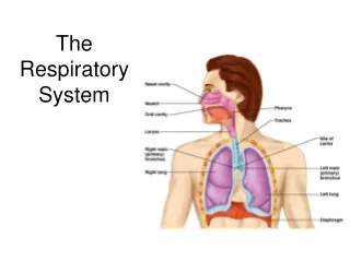
The Respiratory System. The Larynx (voicebox). Extends from the level of the 4 th to the 6 th cervical vertebrae Attaches to hyoid bone superiorly Inferiorly is continuous with trachea (windpipe) Three functions: Produces vocalizations (speech) Provides an open airway (breathing)

E N D
The Larynx (voicebox) • Extends from the level of the 4th to the 6th cervical vertebrae • Attaches to hyoid bone superiorly • Inferiorly is continuous with trachea (windpipe) • Three functions: • Produces vocalizations (speech) • Provides an open airway (breathing) • Switching mechanism to route air and food into proper channels • Closed during swallowing • Open during breathing
Framework of the larynx • 9 cartilages connected by membranes and ligaments • Thyroid cartilage with laryngeal prominence (Adam’s apple) anteriorly • Cricoid cartilage inferior to thyroid cartilage: the only complete ring of cartilage: signet shaped and wide posteriorly
Behind thyroid cartilage and above cricoid: 3 pairs of small cartilages • Arytenoid: anchor the vocal cords • Corniculate • Cuneiform • 9th cartilage: epiglottis
* * Posterior views Epliglottis* (the 9th cartilage) Elastic cartilage covered by mucosa On a stalk attached to thyroid cartilage Attaches to back of tongue During swallowing, larynx is pulled superiorly Epiglottis tips inferiorly to cover and seal laryngeal inlet Keeps food out of lower respiratory tract
Cough reflex: keeps all but air out of airways • Low position of larynx is required for speech (although makes choking easier) • Paired vocal ligaments: elastic fibers, the core of the true vocal cords
Pair of mucosal vocal folds (true vocal cords) over the ligaments: white because avascular
Glottis is the space between the vocal cords • Laryngeal muscles control length and size of opening by moving arytenoid cartilages • Sound is produced by the vibration of vocal cords as air is exhaled
Innervation of larynx (makes surgery at neck risky) • Recurrent laryngeal nerves of Vagus • These branch off the Vagus and make a big downward loop under vessels, then up to larynx in neck • Left loops under aortic arch • Right loops under right subclavian artery • Damage to one: hoarseness • Damage to both: can only whisper
Trachea (the windpipe) • Descends: larynx through neck into mediastinum • Divides in thorax into two main (primary) bronchi • 16-20 C-shaped rings of hyaline cartilage joined by fibroelastic connective tissue • Flexible for bending but stays open despite pressure changes during breathing
Posterior open parts of tracheal cartilage abut esophagus • Trachealis muscle can decrease diameter of trachea • Esophagus can expand when food swallowed • Food can be forcibly expelled • Wall of trachea has layers common to many tubular organs – filters, warms and moistens incoming air • Mucous membrane (pseudostratified epithelium with cilia and lamina propria with sheet of elastin) • Submucosa ( with seromucous glands) • Adventitia - connective tissue which contains the tracheal cartilages)
Carina* • Ridge on internal aspect of last tracheal cartilage • Point where trachea branches (when alive and standing is at T7) • Mucosa highly sensitive to irritants: cough reflex *
Bronchial tree bifurcation • Right main bronchus (more susceptible to aspiration) • Left main bronchus • Each main or primary bronchus runs into hilus of lung posterior to pulmonary vessels 1. Oblique fissure2. Vertebral part3. Hilum of lung4. Cardiac impression5. Diaphragmatic surface (Wikipedia)
Main=primary bronchi divide into secondary=lobar bronchi, each supplies one lobe • 3 on the right • 2 on the left • Lobar bronchi branch into tertiary = segmental bronchi • Continues dividing: about 23 times • Tubes smaller than 1 mm called bronchioles • Smallest, terminal bronchioles, are less the 0.5 mm diameter • Tissue changes as becomes smaller • Cartilage plates, not rings, then disappears • Pseudostratified columnar to simple columnar to simple cuboidal without mucus or cilia • Smooth muscle important: sympathetic relaxation (“bronchodilation”), parasympathetic constriction (“bronchoconstriction”)
Respiratory Zone • End-point of respiratory tree • Structures that contain air-exchange chambers are called alveoli • Respiratory bronchioles lead into alveolar ducts: walls consist of alveoli • Ducts lead into terminal clusters called alveolar sacs – are microscopic chambers • There are 3 million alveoli!
Gas Exchange • Air filled alveoli account for most of the lung volume • Very great area for gas exchange (1500 sq ft) • Alveolar wall • Single layer of squamous epithelial cells (type 1 cells) surrounded by basal lamina • 0.5um (15 X thinner than tissue paper) • External wall covered by cobweb of capillaries • Respiratory membrane: fusion of the basal laminas of • Alveolar wall • Capillary wall Respiratory bronchiole Alveolar duct (air on one side; blood on the other) Alveoli Alveolar sac
Bronchial “tree” and associated Pulmonary arteries
This “air-blood barrier” (the respiratory membrane) is where gas exchange occurs • Oxygen diffuses from air in alveolus (singular of alveoli) to blood in capillary • Carbon dioxide diffuses from the blood in the capillary into the air in the alveolus
Lungs and Pleura Around each lung is a flattened sac of serous membrane called pleura Parietal pleura – outer layer Visceral pleura – directly on lung Pleural cavity – slit-like potential space filled with pleural fluid • Lungs can slide but separation from pleura is resisted (like film between 2 plates of glass) • Lungs cling to thoracic wall and are forced to expand and recoil as volume of thoracic cavity changes during breathing
Pleura also divides thoracic cavity in three • 2 pleural, 1 mediastinal • Pathology • Pleuritis • Pleural effusion
Lungs • Each is cone-shaped with anterior, lateral and posterior surfaces contacting ribs • Superior tip is apex, just deep to clavicle • Concave inferior surface resting on diaphragm is the base apex apex base base
Abbreviations in medicine: e.g.” RLL pneumonia” Horizontal fissure • Right lung: 3 lobes • Upper lobe • Middle lobe • Lower lobe • Left lung: 2 lobes • Upper lobe • Lower lobe Oblique fissure Oblique fissure Each lobe is served by a lobar (secondary) bronchus
Muscles of Inspiration • During inspiration, the dome shaped diaphragm flattens as it contracts • This increases the height of the thoracic cavity • The external intercostalmuscles contract to raise the ribs • This increases the circumference of the thoracic cavity Together:
Inspiration continued • Intercostals keep the thorax stiff so sides don’t collapse in with change of diaphragm • During deep or forced inspiration, additional muscles are recruited: • Scalenes • Sternocleidomastoid • Pectoralis minor • Quadratus lumborum on 12th rib • Erector spinae (some of these “accessory muscles” of ventilation are visible to an observer; it usually tells you that there is respiratory distress – working hard to breathe)
Expiration • Quiet expiration in healthy people is chiefly passive • Inspiratory muscles relax • Rib cage drops under force of gravity • Relaxing diaphragm moves superiorly (up) • Elastic fibers in lung recoil • Volumes of thorax and lungs decrease simultaneously, increasing the pressure • Air is forced out
Expiration continued • Forced expiration is active • Contraction of abdominal wall muscles • Oblique and transversus predominantly • Increases intra-abdominal pressure forcing the diaphragm superiorly • Depressing the rib cage, decreases thoracic volume • Some help from internal intercostals and latissimus dorsi (try this on yourself to feel the different muscles acting)


