Download
1 / 36
460 likes | 1.86k Views
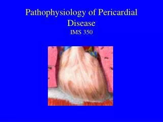
Pathophysiology of Pericardial Disease IMS 350. Pericardium - Anatomy. Normal pericardium is a fibro-serous sac which surrounds the heart and adjoining portions of the great vessels.

E N D
Pericardium - Anatomy • Normal pericardium is a fibro-serous sac which surrounds the heart and adjoining portions of the great vessels. • The inner visceral layer, also known as the epicardium, consists of a thin layer of mesothelial cells closely adherent to the surface of the heart. The epicardium is reflected onto the surface of the outer fibrous layer with which it forms the parietal pericardium. • The parietal pericardium consists of collagenous fibrous tissue and elastic fibrils. • Between the two layers lies the pericardial space, which contains approximately 10-50ml of fluid, which is an ultrafiltrate of plasma. • Drainage of pericardial fluid is via right lymphatic duct and thoracic duct.
Pericardium: Anatomy • Pericardial Layers: • Visceral layer • Parietal layer • Fibrous pericardium
Function of the Pericardium 1. Stabilization of the heart within the thoracic cavity by virtue of its ligamentous attachments -- limiting the heart’s motion. 2. Protection of the heart from mechanical trauma and infection from adjoining structures. 3. The pericardial fluid functions as a lubricant and decreases friction of cardiac surface during systole and diastole. 4. Prevention of excessive dilation of heart especially during sudden rise in intra-cardiac volume (e.g. acute aortic or mitral regurgitation).
Etiologies of Pericarditis I. INFECTIVE 1. VIRAL - Coxsackie A and B, Influenza, adenovirus, HIV, etc. 2. BACTERIAL - Staphylococcus, pneumococcus, tuberculosis, etc. 3. FUNGAL - Candida 4. PARASITIC - Amoeba, candida, etc. II. AUTOIMMUNE DISORDERS 1. Systemic lupus erythematosus (SLE) 2. Drug-Induced lupus (e.g. Hydralazine, Procainamide) 3. Rheumatoid Arthritis 4. Post Cardiac Injury Syndromes i.e. postmyocardial Infarction (Dressler's) Syndrome, postcardiotomy syndrome, etc. III. NEOPLASM 1. Primary mesothelioma 2. Secondary, metastatic 3. Direct extension from adjoining tumor IV. RADIATION PERICARDITIS V. RENAL FAILURE (uremia) VI. TRAUMATIC CARDIAC INJURY 1. Penetrating - stab wound, bullet wound 2. Blunt non-penetrating - automobile steering wheel accident VII. IDIOPATHIC
Pathogenesis 1) Vasodilation: transudation of fluid 2) Increased vascular permeability leakage of protein 3) Leukocyte exudation neutrophils and mononuclear cells Pathology depends on underlying cause and severity of inflammation serous pericarditis serofibrinous pericarditis suppurative (purulent) pericarditis hemorrhagic pericarditis
Clinical Features of Acute Pericarditis Idiopathic/viral * Pleuritic Chest pain * Fever * Pericardial Friction Rub 3 component: a) atrial or pre-systolic component b) ventricular systolic component (loudest) c) ventricular diastolic component * EKG: diffuse ST elevation PR segment depression
Diagnostic Tests Echocardiogram: Pericardial effusion N.B.: absence does not rule out pericarditis N.B.: Pericarditis is a clinical diagnosis, not an Echo diagnosis! Blood tests: PPD, RF, ANA Viral titers Search for malignancy Pericardiocentesis: low diagnostic yield done therapeutically
Treatment Pain relief analgesics and anti-inflammatory ASA/NSAID’s Steroids for recurring pericarditis Antibiotics/drainage for purulent pericarditis Dialysis for uremic pericarditis Neoplastic: XRT, chemotherapy
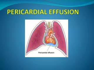
Pericardial Effusion Normal 15-50 ml of fluid • ETIOLOGY • 1. Inflammation from infection, immunologic process. • 2. Trauma causing bleeding in pericardial space. • 3. Noninfectious conditions such as: • a. increase in pulmonary hydrostatic pressure e.g. congestive heart failure. • b. increase in capillary permeability e.g. hypothyroidism • c. decrease in plasma oncotic pressure e.g. cirrhosis. • 4. Decreased drainage of pericardial fluid due to obstruction of thoracic duct as a result of malignancy or damage during surgery. • Effusion may be serous, serofibrinous, suppurative, chylous, or hemorrhagic depending on the etiology. • Viral effusions are usually serous or serofibrinous • Malignant effusions are usually hemorrhagic.
Pathophysiology Pericardium relatively stiff Symptoms of cardiac compression dependant on: 1. Volume of fluid 2. Rate of fluid accumulation 3. Compliance characteristics of the pericardium A. Sudden increase of small amount of fluid (e.g. trauma) B. Slow accumulation of large amount of fluid (e.g. CHF)
Clinical features Small effusions do not produce hemodynamic abnormalities. Large effusions, in addition to causing hemodynamic compromise, may lead to compression of adjoining structures and produce symptoms of: dysphagia (compression of esophagus) hoarseness (recurrent laryngeal nerve compression) hiccups (diaphragmatic stimulation) dyspnea (pleural inflammation/effusion)
Physical Findings Physical Findings: • Muffled heart sounds • Paradoxically reduced intensity of rub • Ewart's sign: Compression of lung leading to an area of consolidation in the left infrascapular region (atalectasis, detected as dullness to percussion and bronchial breathing)
Diagnostic studies • CXR: “water bottle” shaped heart • EKG: • low voltage • “electrical alternans” • Echocardiogram
Cardiac Tamponade Fluid under high pressure compresses the cardiac chambers: acute: trauma, LV rupture – may not be very large gradual: large effusion, due to any etiology of acute pericarditis
CardiacTamponade -- Pathophysiology Accumulation of fluid under high pressure: compresses cardiac chambers & impairs diastolic filling of both ventricles SV venous pressures CO systemic pulmonary congestion Hypotension/shock JVD rales Reflex tachycardia hepatomegaly ascites peripheral edema
Tamponade-- Clinical Features Symptoms: Acute: (trauma, LV rupture) profound hypotension confusion/agitation Slow/Progressive large effusion (weeks) Fatigue (CO) Dyspnea JVD Signs: Tachycardia Hypotension rales/edema/ascites muffled heart sounds pulsus paradoxus
Pulsus Paradoxus • Intrapericardial pressure (IPP) tracks intrathoracic pressure. • Inspiration: • negative intrathoracic pressure is transmitted to the pericardial space • IPP • blood return to the right ventricle • jugular venous and right atrial pressures • right ventricular volume interventricular septum shifts towards the left ventricle • left ventricular volume • LV stroke volume • blood pressure (<10mmHg is normal) during inspiration
Pulsus Paradoxus Exaggeration of normal physiology > 10 mm Hg drop in BP with inspiration
Tamponade -- Diagnosis EKG: low voltage, sinus tachycardia, electrical alternans Echocardiography pericardial effusion (r/o other etiologies in dif dx) RA and RV diastolic collapse
Right Heart Catheterization Catheterization Findings: Elevated RA and RV diastolic pressures Equalized diastolic pressures Blunted “y” descent in RA tracing y descent: early diastolic filling (atrial emptying) BP and Pulsus paradoxus Pericardial pressure = RA pressure
Jugular venous pressure waves • Normal JVP contours (1) A-wave • 1) results from ATRIAL contraction • 2) Timing - PRESYSTOLIC • 3) Peak of the a-wave near S1 (2) V-wave 1) results from PASSIVE filling of the right atrium while the tricuspid valve is closed during ventricular systole (Remember the V-wave is a "V"ILLING WAVE) 2) Large V-waves on the left side of the heart may be seen with mitral regurgitation, atrial septal defect, ventricular septal defect. The v-wave in the jugular venous pulse reflects right atrial events. To see the v-wave on the left side of the heart Swan-Ganz monitoring is needed 3) timing - peaks just after S2 (3) X-descent 1) results from ATRIAL RELAXATION 2) timing - occurs during ventricular systole, at the same time as the carotid pulse occurs (4) Y-descent 1) results from a FALL in right atrial pressure associated with opening of the tricuspid valve 2) timing - occurs during ventricular diastole (5) Generalizations 1) the A-wave in a normal individual is always larger than the V-wave 2) the X-descent is MORE PROMINENT than the Y-descent
RA Pressure Tracing a wave: atrial contraction v wave: passive filling of atria during ventricular systole with mv/tv closed y descent: early atrial emptying with mv/tv open (early passive filling of ventricle) Tamponade: blunted y descent (impaired rapid ventricular filling due to compression by high pericardial pressure)
Tamponade -- Treatment Pericardiocentesis Pericardial Window Balloon Pericardiotomy Pre-pericardiocentisis Post-pericardiocentesis
Constrictive Pericarditis Late complication of pericardial disease Fibrous scar formation Fusion of pericardial layers Calcification further stiffens pericardium Etiologies: any cause of pericarditis idiopathic post-surgery tuberculosis radiation neoplasm
Pathophysiology Rigid, scarred pericardium encircles heart: Systolic contraction normal Inhibits diastolic filling of both ventricles SV venous pressures CO systemic pulmonary congestion Hypotension/shock JVD rales Reflex tachycardia hepatomegaly ascites peripheral edema
Physical exam HR, BP ascites, edema, hepatomegaly early diastolic “knock” after S2 sudden cessation of ventricular diastolic filling imposed by rigid pericardial sac Kussmaul’s sign
Kussmaul’s Sign inspiration: intrathoracic pressure, venous return to thorax intrathoracic pressure not transmitted though to RV no pulsus paradoxus! no inspiratory augmentation of RV filling (rigid pericardium) intrathoracic systemic veins become distended JVP rises with inspiration (normally falls)
Diagnosis CXR: calcified cardiac silhouette EKG: non-specific CT or MRI: pericardial thickening
Cardiac Catheterization Elevated and equalized diastolic pressures (RA=RVEDP=PAD=PCW) Prominent y descent: “dip and plateau”: rapid atrial emptying rapid ventricular filling then abrupt cessation of blood flow due to rigid pericardium
Constriction vs. Restriction Similar presentation and physiology, important to differentiate as constriction is treatable by pericardiectomy Majority of diseases causing restriction are not treatable
Constrictive Pericarditis Tachycardia, low voltage Equalized diastolic pressures Thickened pericardium Thickened pericardium RV=LV,dip & plateau Kussmaul’s
TAMPONADE Low cardiac output state JVD present NO Kussmaul’s sign Equalized diastolic pressures RA: blunted y descent Decreased heart sounds CONSTRICTION Low cardiac output state JVD present Kussmaul’s sign Equalized diastolic pressures RA: rapid y descent Pericardial “knock” Constriction vs. Tamponade Summary
TAMPONADE Pulsus paradoxus: Present Echo/MRI: Normal systolic function Large effusion RA & RV compression Treatment: Pericardiocentesis CONSTRICTION Pulsus paradoxus: Absent Echo/MRI: Normal systolic function No effusion Pericardial thickening Treatment: Pericardial stripping Constriction vs. Tamponade Summary