Download

1 / 8
90 likes | 232 Views
‘How I do’ the Evaluation of Pericardial Disease. Brian J. Schietinger MD and Christopher M. Kramer MD For scmr.org From the University of Virginia

E N D
‘How I do’ the Evaluation of Pericardial Disease Brian J. Schietinger MD and Christopher M. Kramer MD For scmr.org From the University of Virginia This presentation posted for members of scmr as an educational guide – it represents the views and practices of the author, and not necessarily those of SCMR. bjs3p@virginia.edu
1. Multi-Slice Localizers Coronal Sagittal Transverse • • Pericardial disorders often distort normal cardiac anatomy. Therefore, obtaining classic views may require careful image planning. • Note: Pericardial diseases have characteristic anatomical features, but the diagnosis remains a clinical and hemodynamic one.
2. SSFP Cine Pericardial Effusion Effusive Constrictive Pericarditis • Obtain steady state free precession (SSFP) cine images in two, three, and four chamber cardiac orientations. • Above are four chamber SSFP cine views of representative pericardial disorders. Pericardial Constriction
3. Stacked Axial SSFP Cine • Stacked axial SSFP cines are useful to determine pericardial regions of interest and the extent of involvement.
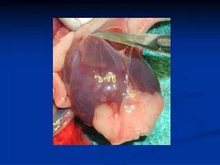
4. T1W and T2W spin echo imaging: Pericardial Cysts 2 1 T1W Spin Echo T2W Spin Echo • Pericardial cysts (white arrows) often are located at the right diaphragmatic border and are characterized with T1 weighted and T2 weighted turbo spin echos (TSE). • T1 weighted TSE sequences (Image 1) demonstrate hypointense (dark) signal in the pericardial cyst. • T2 weighted TSE sequences (Image 2) demonstrate hyperintense (bright) signal in the pericardial cyst which contains proteinaceous fluid.
5. T1W TSE and fat suppressed gradient echo imaging: Constrictive pericarditis 2 1 • Correctly identifying the pericardium requires that it be delineated from fat and fluid. To do so often is aided by acquiring both a T1 weighted TSE and a T1 weighted fat suppressed gradient echo (GRE) sequence. • T1 weighted TSE sequences (Image 1) produce hyperintense (bright) epicardial and pericardial fat (yellow arrows), hypointense (dark) pericardium (white arrow), and, if present, hypointense (dark) pericardial fluid. No pericardial fluid is seen in this case. • Note: Normal pericardium is ≤ 3mm thick. • T1 weighted fat suppressed GRE sequence (Image 2) nulls signal from epicardial (yellow arrow) and pericardial fat. In contrast, the pericardium appears bright (white arrow).
6. GRE tagged imaging: Constrictive pericarditis • Myocardial tagging is integral to the determination of pericardial adherence to the epicardium. Normal pericardium should slip over the epicardial surface. Note: Fluid and fat do not “hold” tags. • Often a left ventricular short axis tagged cine in the atrioventricular groove can be very helpful for identifying the presence and extent of pericardial constriction. Constrictive Pericarditis EffusiveConstrictive Pericarditis