Pericardial Disease: Causes, Symptoms & Management
330 likes | 423 Views
Learn about Acute and Chronic Pericarditis, Constrictive Pericarditis, Cardiac Tamponade, and more. Explore pericardial anatomy, physiology, inflammation, and pathology. Discover diagnostic clues, ECG features, and management strategies.

Pericardial Disease: Causes, Symptoms & Management
E N D
Presentation Transcript
Pericardial DiseaseBy Dr. Muhammad Aftab ShahSenior Registrar CardiologyKEMU/Mayo Hospital, Lahore.
Pericardial Disease • Acute Pericarditis • Chronic Relapsing Pericarditis • Constrictive Pericarditis • Cardiac Tamponade • Localized and Low Pressure Tamponade • Restrictive Cardiomyopathy
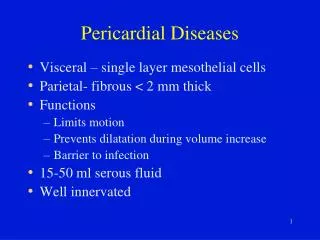
Pericardial Anatomy • Two major components • serosa (viceral pericardium)mesothelial monolayerfacilitate fluid and ion exchange • fibroa (parietal pericardium)fibrocollagenous tissue • Pericardial Fluid • 15 - 50 ml of clear plasma ultrafiltrate • Ligamentous attachments • to the sternum, vertebral column, diaphragm
Pericardial Physiology • not needed to sustain life • physiologic functions • limit cardiac dilatation • maintain normal ventricular compliance • reduce friction to cardiac movement • barrier to inflammation • limit cardiac displacement
Pericardial Inflammationpathogenesis • Contiguous spread • lungs, pleura, mediastinal lymph nodes, myocardium, aorta, esophagus, liver • Hematogenous spread • septicemia, toxins, neoplasm, metabolic • Lymphangetic spread • Traumatic or irradiation
Pericardial Inflammationpathology • inflammation provokes a fibrinous exudate with or without serous effusion • the normal transparent and glistening pericardium is turned into a dull, opaque, and “sandy” sac • can cause pericardial scarring with adhesions and fibrosis
Acute Pericarditiscommon causes • Outpatient setting • usually idiopathic • probably due to viral infections • Coxsackie A and B (highly cardiotropic) are the most common viral cause of pericarditis and myocarditis • Others viruses: mumps, varicella-zoster, influenza, Epstein-Barr, HIV
Acute Pericarditiscommon causes • Inpatient settingT = Trauma, TUMORU = UremiaM = Myocardial infarction (acute, post) Medications (hydralazine, procain)O = Other infections (bacterial, fungal, TB)R = Rheumatoid, autoimmune disorder Radiation
Acute PericarditisDiagnostic Clues • History sudden onset of anterior chest pain that is pleuritic and substernal • Physical exam presence of two- or three-component rub • ECG most important laboratory clue
Chest Pain Historypericarditis vs infarction • Common characteristics • retrosternl or precordial with raditaion to the neck, back, left shoulder or arm • Special characteristics (pericarditis) • more likely to be sharp and pleuritic • with coughing, inspiration, swallowing • worse by lying supine, relieved by sitting and leaning forward
Heart Murmurs of Pericarditis • Pericardial friction rub is pathognomic for pericarditis • scratching or grating sound • Classically three components: • presystolic rub during atrial filling • ventricular systolic rub (loudest) • ventricular diastolic rub (after A2P2)
Acute PericarditisECG features • ST-segment elevation • reflecting epicardial inflammation • leads I, II, aVL, and V3-V6 • lead aVR usually shows ST depression • ST concave upward • ST in AMI concave downward like a “dome” • PR segment depression (early stage) • T-wave inversion • occurs after the ST returns to baseline
Acute PericarditisManagement • Treat underlying cause • Analgesic agents • codeine 15-30 mg q 4-6 hr • Anti-inflmmatory agents • ASA 648 mg q 3-4 hrs • NSAID (indomethacin 25-50 mg qid) • Corticosteroids are symptomatically effective , but preferably avoided
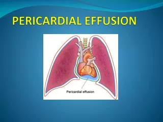
Types of Effusive Fluid • serous • transudative - heart failure • suppurative • pyogenic infection with cellular debris and large number of leukocytes • hemorrhagic • occurs with any type of pericarditis • especially with infections and malignancies • serosanguinous
Dignostic Evaluation • Chest x-ray • usually requires > 200 ml of fluid • cannot distinguish between pericardial effusion and cardiomegly • Echocardiography • standard for diagnosing pericardial effusion • convenient, highly reliable, cost effective • false positives (M-mode)- left pleural effusion, epicardial fat, tumor tissue, pericardial cysts
Noncompressing Effusion • asymptomatic unless they are large enough to compress adjacent organs • dysphagia • cough • dyspnea • hoarseness • hiccups • abdminal fullness • nausea
Cardiac Tamponade • Decompensated cardiac compression from increased intracardaic press
Cardiac Tamponade • Early stage • mild to moderate elevation of central venous pressure • Advanced stage • intrapericardial pressure ventricular filling, stroke volume • hypotension • impaired organ perfusion
Beck’s Triad • Described in 1935 by thoracic surgeon Claude S. Beck • 3 features of acute tamponade • Decline in systemic arterial pressure • Elevation in systemic venous pressure (e.g. distended neck vein) • A small, quiet heart
Cardiac TamponadeBedside Diagnosis • Elevated jugular venous pressure • Paradoxical pulse
Pulsus Paradoxus • an exaggerated drop in blood pressure with inspiration (>10mmHg) • tamponade without pulsus • atrial septal defect • aortic insufficiency • LVH with LVEDP • pulsus without tamponade • COPD, RV infarct, pulmonary embolism
Echocardiography • Pericardial effusion • highly reliable • Cardiac tamponade • RA and RV diastolic collapse • reduced chamber size • distension of the inferior vena cava • exaggerated respiratory variation of the mitral and tricuspid valve flow velocities
Pericardiocentesis • Diagnostic tap • usually not indicated • rarely have positive cytology or infection that can be diagnosed • Therapeutic drainage • indicated for significant elevation of the central venous pressure