Download
1 / 69
710 likes | 1.47k Views
Haemodynamics of pericardial diseases. DEEPAK NANDAN. Pericardium - Anatomy. F ibro-serous sac The inner visceral layer- - thin layer of mesothelial cells.

E N D
Haemodynamics of pericardial diseases DEEPAK NANDAN
Pericardium - Anatomy • Fibro-serous sac • The inner visceral layer-- thin layer of mesothelial cells. • Parietal pericardium- collagenous fibrous tissue and elastic fibrils. • Between the 2layers lies the pericardial space- 10-50ml of fluid-ultrafiltrateof plasma. • Drainage of pericardial fluid is via right lymphatic duct and thoracic duct.
Pericardium: Anatomy • Pericardial Layers: • Visceral layer • Parietal layer • Fibrous pericardium
1)Effects on chambers Limits short-term cardiac distention Facil chamber coupling and diast interaction Maint P-V relation of chambers and output Maint geometry of left ventricle 2) Effects on whole heart Lubricates, min friction Equal gravit inertial, hydrostatic forces 3) Mech barrier to infection 4) Immunologic 5) Vasomotor 6) Fibrinolytic 7) Modulation of myo structure and function and gene expression
Physiology of the Pericardium • Limits distension of the cardiac chambers • Facilitates interaction and coupling of the ventricles and atria. • Changes in pressure and volume on one side of the heart can influence pressure and volume on the other side • Influences quant and qualit aspects of vent filling- RV and RA > influence of the pericardium than is the resistant, thick-walled LV.
Magnitude & imp of pericardial restraint of vent filling at physiologic cardiac volumes- controversial • Pericardial reserve volume - diff between unstressed pericardial volume and cardiac volume. • PRV-relatively small & peri influences become signi when the reserve volume is exceeded • Rapid ↑ in blood volume • Rapid ↑ in heart size-a/c acuteMR, pulm embolism, RV infarction
Stress-strain and pressure-volume curves of the normal pericardium.
Flat compliant segment transitions abruptly to noncompliant seg • Small reserve volume –exceeded , pr within the sac –acting on the heart ↑ rapidly-transmitted to inside the chambers • Once critical level of effusion is reached- small amounts of addl fluid –marked ↑ peri pr and ↓ function • Removal of small amounts- improves
Chronic stretching of the pericardium results in "stress relaxation“ • Large but slowly developing effusions do not produce tamponade. • Pericardium adapts to cardiac growth by "creep" (i.e., an increase in volume with constant stretch) and cellular hypertrophy
Restrain cardiac vol • Force it exerts on the heart influences filling • A component of intracavitary filling pressure –transmission of peri pr • Contact pr is more imp 4 R heart which have a lower filling pressure than L • Diastolic interaction • Transmission of intracavitary pr to adjoining chambers • Once card vol↑ above phy range-pericardium contributes ↑nly to filling pressure dir-contact pr indir-diastolic interaction
3 possible pericardial compression syndromes Cardiac tamponade • Accumulation of pericardial fluid under pressure and may be acute or subacute Constrictive pericarditis • Scarring and consequent loss of elasticity of the pericardial sac Effusive-constrictive pericarditis • Constrictive physiology with a coexisting pericardial effusion
CardiacTamponade -- Pathophysiology Accumulation of fluid under high pressure: compresses cardiac chambers & impairs diastolic filling of both ventricles SV venous pressures CO systemic pulmonary congestion Hypotension/shock ↑JVPrales Reflex tachycardia hepatomegaly ascites peripheral edema
Pathophysiology Pericardium relatively stiff Symptoms of cardiac compression dependant on: 1. Volume of fluid 2. Rate of fluid accumulation 3. Compliance characteristics of the pericardium A. Sudden increase of small amount of fluid (e.g. trauma) B. Slow accumulation of large amount of fluid (e.g. CHF)
↑intrapericardial pr-throughout the cardiac cycle-> ↓ cardiac vol during ejection- momentary relief • Nl –biphasic venous return- at the vent ejection - early diastole-TV opens • In tamponade– unimodal - vent systole • Severe tamp- venous return halted in diastole-when cardiac vol & peri pr are maximal • ↓intrathoracic pr in inspiration is transmitted to heart- preserved venous return- kussmaul absent
Hemodynamic features of Cardiac Tamponade • Decrease in CO from reduced SV + increase in CVP • Equalization of diastolic pressure throughout the heart RAP=LAP=RVEDP=LVEDP • Reduced transmural filling pr • Total cardiac volume relatively fixed-small • Blood enters only when blood leaves the chamber --CVP waveform accentuated x descent + abolished y descent
As the fluid accumulates in the peri sac-L&R sided pr rises and equalises to a pr llar to that of peri pressure(15-20mm) • Closest during inspiration • Vent filling press decided by the pr in pericardial sac- prog decline in the EDV • Compensatory ↑ in contractility & heart rate-↓ESV • Not sufficient to normalise SV-CO↓
Absence of Y Descent Wavein Cardiac Tamponade • Bcoz- equalization of 4chambers pressures, no blood flow crosses the atrio-ventricular valve in early diastole (passive ventricular filling, Y descent) • X wave occurs during ventricular systole-when blood is leaving from the heart-prominent
PulsusParadoxus • Intraperipressure (IPP) tracks- intrathoracic pressure. • Inspiration: • -veintrathoracicpressure is transmitted to the pericardial space • IPP • blood return to the right ventricle • jugular venous and right atrial pressures • right ventricular volume IVS shifts towards the left ventricle • left ventricular volume • LV stroke volume • blood pressure (<10mmHg is normal) during inspiration
PulsusParadoxus > 10 mm Hg drop in BP with inspiration Exaggeration of normal physiology
Other factors ↑afterload –transmission of-veintrathoracic pr to aorta Traction on the pericardium caused by descent of the diaphragm-↑ peric pr Reflex changes in vas resistance& card contractility ↑ respi effort due to pulmonary congestion
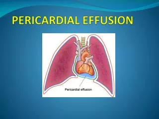
Pericardial tamponade after pericardiocentesis
Stress Responses to Cardiac Tamponade • Reflex sympathetic activation => ↑HR + contractility • Arterial vasoconstriction to maintain systemic BP • Venoconstrictionaugments venous return • Relatively fixed SV • CO is rate dependent
TAMPONADE WITHOUT PP • When preexisting elevations of diastolic pressures/ volumes exist –no PP • Eg;- LV dysfunction AR ASD Aortic dissection with AR
Low pressure tamponade • Intrvascular volume low in a preexisting effusion • Modest ↑ in peri pr can compromise already↓ SV • Dialysis patient • Diuretic to effusion patient • Pats with blood loss and dehydration • JVP & pulsusparadoxus absent
Pathophysiology Rigid, scarred pericardium encircles heart: Systolic contraction normal Inhibits diastolic filling of both ventricles SV venous pressures CO systemic pulmonary congestion Hypotension/shock↑ JVPrales Reflex tachycardia hepatomegaly ascites peripheral edema
Pathophysiology Heart encased by rigid ,non compliant shell 1. uniform impairment of RV and LV filling EARLY DIASTOLIC filling normal(↑RAP+suction due to ↓ESV) filling abruptly halted in mid and late diastole pressure rises mid to late diastole 2. ↑interventricular interdependence 3. dissociation of thoracic and cardiac chambers - Kussmaul’s - decreased LV filling with inspiration and increased RV filling
CP- card vol is fixed- attained after initial1/3rd of diastole • Biphasic venous return- dias≥ to systolic component • Card compression insignificant –end systole + • ↑RAP+vent suction due to ↓ ESV- rapid early diastolic filling
Kussmaul’s Sign Inspiration: intrathoracicpr, venous return to thorax intrathoracicpr not transmitted to RV no pulsusparadoxus no inspiratory augmentation of RV filling (rigid pericardium) intrathoracic systemic veins become distended JVP rises with inspiration
Kussmaul’s Sign • Mechanism: 1) Increase venpressure due to ↓compliance of pericardium and heart2) ↑abdominal presssure during inspiration with elevated venous pressure • Clinical presentation: inspiratory engorgementof jugular vein • Also seen in cardiomyopathy, pulmonaryembolism, and RVMI
Friedreich's sign • Early diastolic pressure dip observed in cervical veins or recorded from RA / SVC • Rapid early filling of vent-↑ RAP+ suction due to ↓ ESV
HEMODYNAMICS OF CP • Impairement of RV/LV filling with chamber vol limited by rigid pericardium 1) high RAP with prom X & Y descent 2) ‘Squre root’ sign of RV & LV PR wave form 3) PASP & RVSP < 50 mm Hg 4) RVEDP> 1/3 RVSP • ↑Interventricular dependence & dissociation of thoracic & cardiac chambers 1) kussmaul’s sign 2) RVEDP & LVEDP < 5 mm apart 3) Respiratory discordance in peak RVSP & LVSP
↓intra thoracic pr fails to get transmitted into heart- inspirat ↑ in venous return doesn’t occur- Kussmaul’s sign • Inspiratory ↑ in ven return & RV vol-doesn’t occur + position of vent septum not dramatically altered =no pulsusparadoxus
Cath • ↑ RAP • Prominent X and Y descents of atrial pressure tracings • ↑RVEDP ≥ 1/3 of RVSP • "Square root" signs in the RV and LV diastolic pressure tracings • >insp↓in PCWP compared to LVEDP • Equalization of LV and RV diastolic plateau pressure tracings • Discordance between RV and peak LV systolic pressures during inspiration(100%sen,spec)
Cardiac Catheterization Elevated and equalized diastolic pressures (RA=RVEDP=PAD=PCW) Prominent y descent: “dip and plateau”: rapid atrial emptying rapid ventricular filling then abrupt cessation of blood flow due to rigid pericardium