Pericardial Diseases
990 likes | 1.88k Views
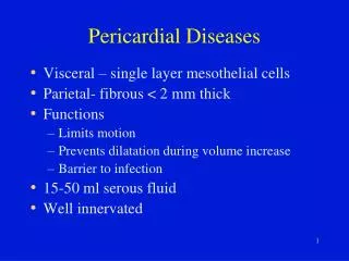
Pericardial Diseases. Israel Freeman, MD Associate Professor of Clinical Medicine. Diseases of the Pericardium. Acute Pericarditis Pericardial Effusion Constrictive Pericarditis. Acute Pericarditis. Acute Pericarditis common causes. Outpatient setting usually idiopathic

Pericardial Diseases
E N D
Presentation Transcript
Pericardial Diseases Israel Freeman, MD Associate Professor of Clinical Medicine
Diseases of the Pericardium • Acute Pericarditis • Pericardial Effusion • Constrictive Pericarditis
Acute Pericarditiscommon causes Outpatient setting • usually idiopathic • probably due to viral infections • Coxsackie A and B (highly cardiotropic) are the most common viral cause of pericarditis and myocarditis • Others viruses: mumps, varicella-zoster, influenza, Epstein-Barr, HIV
Diseases of the Pericardium Acute Pericarditis • Primary: • Usually due to viral infection Course brief and usually uncomplicated • Secondary • Result from other conditions: rheumatic or collagen diseases, radiotherapy or rend failure related, often exhibit clinical feature, similar to acute pericarditis
Acute Pericarditiscommon causes In-patient settingT = Trauma, TUMORU = UremiaM = Myocardial infarction (acute, post) Medications (hydralazine, procainamide)O = Other infections (bacterial, fungal, TB)R = Rheumatoid, autoimmune disorder, Radiation
Special Ethiologic Forms of acute Pericarditis and Pericardial Effusion • Renal failure and dialysis: more extensive dialysis and NSAID is usually successful occasionally pericardocentesis necessary • unexpected drop of B.P during dialysis may be the clue • Radiation induced pericardial effusion: • After radiation to the mediastinum • May evolve into constrictive Pericarditis after many years • Neoplastic pericardial effusion • Accounts for 50% of patient with tamponade • Lung, Breast cancers accounts for majority of causes, lymphomas and leukemia also important causes • Purulent Pericarditis: usually requires surgical draming • Drug induced Pericarditis: Procainamide, Minoxidil, Methylsergide • Post Cardiotomy Syndrome
Acute PericarditisDiagnostic Clues • History sudden onset of anterior chest pain that is pleuritic and substernal • Physical exam presence of two- or three-component rub • ECG most important laboratoryclue
Acute PericarditisDifferential Diagnosis • Acute myocardial infarction • Pulmonary embolism • Pneumonia • Aortic dissection
Acute PericarditisChest Pain Historypericarditis vs infarction • Common characteristics • retrosternl or precordial with raditaion to the neck, back, left shoulder or arm • Special characteristics (pericarditis) • more likely to be sharp and pleuritic • with coughing, inspiration, swallowing • worse by lying supine, relieved by sitting and leaning forward
Acute Pericarditis Heart Murmurs of Pericarditis • Pericardial friction rub is pathognomic for pericarditis • scratching or grating sound • Classically three components: • presystolic rub during atrial filling • ventricular systolic rub (loudest) • ventricular diastolic rub (after A2P2
Acute Pericarditis ECG in stage 1 of pericarditis shows PR segment depression and ST elevations.
Stage PR segments ST segments T waves 1 Depressed Elevated Upright 2 Isoelectric Isoelectric Flat 3 Isoelectric Isoelectric Inverted 4 Isoelectric Isoelectric Upright Stages of ECG Evolution in Acute Pericarditis
ECG in Acute Pericarditis EKG contrast the pattern of ST segment elevation characteristic of acute pericarditis (a) with the normal variant (early repolarization) pattern of ST segment elevation (b). The normal variant pattern is associated with a normal or slow heart rate and has relatively tall R waves and T waves in V4, V5, and V6. The ST segment elevation is less than 25% of the T wave amplitude. In contrast, the acute pericarditis has PR depression and lower T wave.
Acute Pericarditis ECG features • ST-segment elevation • reflecting epicardial inflammation • leads I, II, aVL, and V3-V6 • lead aVR usually shows ST depression • ST concave upward • ST in AMI concave downward like a “dome” • PR segment depression • early stage • T-wave inversion • occurs after the ST returns to baseline
Pericarditis Acute apical infarction Early Repolarization
Acute PericarditisManagement • Analgesic agents • codeine 15-30 mg q 4-6 hr • Anti-inflmmatory agents • Treat underlying cause • ASA 648 mg q 3-4 hrs • NSAID (indomethacin 25-50 mg qid) • Corticosteroids are symptomatically effective , but preferably avoided
Acute Pericarditis • Diagnosis • Chest pain • Friction rub, fever, Tachycardia • EKG changes are common (diffuse ST-T changes with absence of reciprocal ST depression when ST elevation is present, depressed PR segment) • Mild elevation of cardiac enzymes • Treatment • Analgesics – codeine, hydrocodone • NSAID – Indomethacin (100 mg/d) • Corticosteroids – (40 – 60 mg/d) , slow tapering (2 – 4 weeks) due to possible recurrence after steroid withdrawal
Other forms of Acute Pericarditis • Relapsing Pericarditis • Acute Pericarditis of any etiology may relapse • Treatment: Colchicine, Prednisone or immnosuppressants • Progression to constriction • Rarely acute pericarditis progresses to subucute / chromic
Other Forms of PericarditisDressler’s Syndrome • Described by Dressler in 1956 • fever, pericarditis, pleuritis(typically with a low grade fever and a pericardial friction rub) • occurs in the first few days to several weeks following MI or heart surgery • incidence of 6-25% • treat with high-dose aspirin
Other Forms of PericarditisBacterial Pericarditis • Rare condition in antibiotic era (steadily decreased over last 40 years) • Typically arises from contiguous spread of intrathoracic infection (pneumonia, empyema, mediastinitis, endocarditis, trauma, surgery) • Usually fatal without adequate treatment • Diagnosis frequently missed • Often lacks characteristic features of acute pericarditis
Other Forms of PericarditisPurulent Pericarditis(data from 15 patients) # 3 2 3 • mediastinitisesophageal tear, dental abscessretropharyngeal abscess, chest trauma,post-cardiac surgery infection • endocarditis, prosthetic aortic valve • pneumonia (with empyema) • hepatic abscess (with empyema) • bacterial meningitis • diabetes, leukemia, bone marrow transplant 1 1 1 1 3 Arch Int Med 1996; 156:1857
Other Forms of PericarditisTuberculous Pericarditis • Incidence of pericarditis in patients with pulmonary TB ranged from 1-8% • Physical findings: fever, pericardial friction rub, hepatomegaly • TB skin test usually positive • Fluid smear for TB often negative • Pericardial biopsy more definitive
Acute Pericarditis Key Points • Acute pericarditis is accompanied by pericardial effusion in 60% of cases and tamponade in as many as 15% of cases. • Outpatient management of acute idiopathic pericarditis is acceptable if there is no effusion at predischarge echocardiography and surveillance can be provided at home. • In the care of patients in clinically stable condition who are being hospitalized, echocardiography should be performed in the first 24 hours because a large effusion is the best predictor of poor outcome
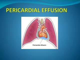
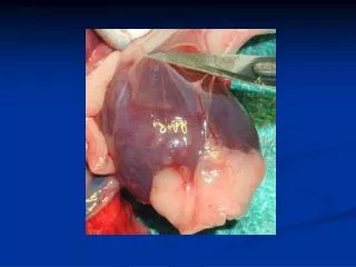
Pericardial Effusion and Cardiac Tamponade • Fluid can accumalte in the pericardium in any form of pericardial disease • Transudate • Exudate • Serosanguineous: Idopathic, dialysis-related, neoplastic, radiation induced, TB, coagulapaties • Bloody: Coagulopathies, trauma, rupture in MI and in aortic dissection • Cardiac Tamponade is the most important complication of fluid accumulation
Cardiac Tamponadepathophysiology Fluid accumulation within the pericardial space resulting in • increased intracardiac pressure • progressive limitation of ventricular diastolic filling • reduction of stroke volume and cardiac output
Cardiac Tamponade • The physiological effect depends on the rate of fluid accumulation • Gradually the pericardium may strech and accommodate > 2,000 ml • Acutely as little as 200 ml of accumulated fluid may raise the intrapercardial pressure to cause tamponade • JVD elevated (rarely not in low pressure tamponade) • B.P variable, paradoxical pulse (>10 mm HG ) • Echocardiography • Treatment: fluids, pericardiocentesis, cardiac surgery.
Cardiac TamponadeBeck’s Triad • Described in 1935 by thoracic surgeon Claude S. Beck • 3 features of acute tamponade • Decline in systemic arterial pressure • Elevation in systemic venous pressure (e.g. distended neck vein) • A small, quiet heart
malignancy idiopathic pericarditis uremia acute myocardial infarction diagnostic procedures with cardiac perforation bacterial tuberculosis radiation myxedema dissecting aortic aneurysm post pericardiotomy syndrome systemic lupus erythematosus cardiomyopathy Etiologies of Cardiac Tamponade
Cardiac Tamponade • Early stage • mild to moderate elevation of central venous pressure • Advanced stage • intrapericardial pressure ventricular filling, stroke volume • hypotension • impaired organ perfusion
Cardiac TamponadeClinical Features • Symptoms • dyspnea, fatigue, agitation and restlessness, syncope, shock, anuria • Physical examination • pulsus paradoxus • tachycardia • increased jugular venous pressure • hypotension
tamponade without pulsus atrial septal defect severe aortic stenosis aortic insufficiency left ventricular dysfunctionLVH with LVEDP decreased intravascular volume (low-pressure tamponade) pulsus without tamponade COPD RV infarct pulmonary embolism effusive constrictive pericarditis restrictive cardiomyopathy extreme obesity tense ascites Cardiac Tamponade Pulsus Paradoxus
Cardiac Tamponade Central Venous Pressure • X - descent • descent of the base in systole • Y - descent • occurs as the tricuspid valve opens and ventricular filling begins from the high-pressure right atrium • in constrictive pericarditis, filling is truncated in early to mid diastole • in tamponade, filling is restricted throughout diastole • Kussmaul’s Sign • in constriction, venous return increases with inspiration and a high right atrial pressure resists filling resulting in an increased JVP
Central Venous Pressure Cardiac Tamponade Constrictive Pericarditis presence of a rapid Y-descent argues against cardiac tamponade
Cardiac Tamponade Pulsus Paradoxus an exaggerated drop in SBP with inspiration (>10mmHg)
Pressure Readings in Cardiac Tamponade The aortic pressure and right atrial pressure are recorded during quiet breathing in a patient with cardiac tamponade. The marked fall in arterial pressure that occurs during inspiration is a paradoxical pulse. The decrease in pulse pressure, defined as the difference between systolic and diastolic pressures, that accompanies the fall in systolic pressure indicates that left ventricular stroke volume decreases during inspiration. Central venous pressure, as indicated by the right atrial pressure, also falls during inspiration.
Cardiac Tamponade Dignostic Evaluation • Chest x-ray • usually requires > 200 ml of fluid • cannot distin guish between pericardial effusion and cardiomegly • Echocardiography • standard for diagnosing pericardial effusion • convenient, highly reliable, cost effective • false positives (M-mode)- left pleural effusion, epicardial fat, tumor tissue, pericardial cysts
ECG in Pericardial Effusion • Diffuse low voltage • amount of fluid • electrical conductivity of the fluid • Electrical alternans • alternating amplitude of the QRS • produced by heart swinging motion • also seen in PSVT, HTN, ischemia
ECG: Pericardial Effusion The electrocardiogram (V2 lead) from a patient with pericardial effusion caused by malignant melanoma reveals a low voltage and electrical alternans.
Cardiac Tamponade Echocardiographic Diagnosis • Pericardial effusion • highly reliable • Cardiac tamponade • RA and RV diastolic collapse • reduced chamber size • distension of the inferior vena cava • exaggerated respiratory variation of the mitral and tricuspid valve flow velocities
Pericardial Effusion-Echocardiography Pericardial effusion is seen in the two-dimensional echocardiogram as an echo-free space outside the cardiac chambers. Two characteristic sites are lateral to the left ventricle in the apical four-chamber view (shown here) and posterior to the left ventricle in the parasternal long-axis view (see Figure 3b).
RA and RV diastolic collapse early diatole systole late diastole
CT Scan: Pericardial Effusion A chest CT scan of a patient with pericardial effusion, pericardial fluid appears less dense than the heart and is separated from the myocardium by epicardial fat in some areas.
Echocardiogram: Pericardial Effusion The echocardiogram demonstrates that the heart moves forward (F) and backward (B) within the effusion on alternate beats, thus producing the alternation of the QRS axis characteristic of electrical alternans. The heart also moves with inspiration (Insp) and expiration (Exp), which accounts for a change in anterior wall motion with every two cardiac cycles.
Differential Diagnosis of Cardiac Tamponade vs. Chronic Constrictive Pericarditis Differences between the central venous pulse contours characteristic of cardiac tamponade (left) and chronic constrictive pericarditis (right) provide the basis for differential diagnosis. The pressure contour in a patient with pericardial effusion and tamponade has a prominent systolic dip (X) but little or no diastolic deflection. The central venous pulse pattern in a patient with chronic constrictive pericarditis displays an M or W contour consisting of both systolic dip (X) and diastolic dip (Y), the Y descent being more prominent.