Download
1 / 14
160 likes | 491 Views
Basics of protein structure and stability III: Anatomy of protein structure. Biochem 565, Fall 2007 08/29/08 Cordes. The backbone conformation of proteins: combination of regular and irregular structures. human hexokinase type I (1QHA) 102 kDa Rosano et al. Structure 1999.

E N D
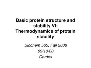
Basics of protein structure and stability III: Anatomy of protein structure Biochem 565, Fall 2007 08/29/08 Cordes
The backbone conformation of proteins:combination of regular and irregular structures human hexokinase type I (1QHA) 102 kDa Rosano et al. Structure 1999. pig insulin (1ZNI) 5.7 kDa Bentley et al. Nature 1976. These “ribbon” diagrams show the “skeleton” of a protein. They are a smoothed representation of the “backbone” or “main chain” structure and do not show the side chains
Backbone or main-chain conformation There are three bonds between “main chain” atoms (everything but the side chain) per residue, and torsional rotation can occur about any of these bonds, in principle. Hence, each residue has 3 angles that describe the main chain conformation for that residue.
Backbone conformation:resonance forms of the peptide group The delocalization of the lone pair of the nitrogen onto the carbonyl oxygen shown in the resonance form on the right imparts significant double bond character (40%) to the peptide bond. Breaking of this double bond character by rotation of the peptide bond requires on the order of 18-21 kcal/mol. Consequently there is not free rotation around the peptide bond: rotation about the peptide bond happens on the time scale of seconds/minutes--very slow
Consequences of double bond character in the peptide bond the peptide C-N bond is 0.12A shorter than the Calpha-N bond. and the C=O is 0.02A longer than that of aldehydes and ketones. All six of the atoms highlighted at left lie in the same plane, and as with carbon-carbon double bonds there are two configurations--cis and trans (trans shown at left)
Consequences of double bond character in the peptide bond trans peptide bond cis peptide bond Still another consequence: in the cis form, the R groups in adjacent residues tend to clash. Hence almost all peptide bonds in proteins are in the trans configuration.
...and that means that the dihedral angle describing rotation around the peptide bond, defined by the four atoms Ca(i)-C-N-Ca(i+1), will generally be close to 180°. This angle is known by the greek symbol w. So the properties of the peptide bond place a strong restriction on the backbone conformation or main-chain conformation of proteins, that is to say, the spatial configuration of the non side-chain atoms.
The peptidyl-proline bond cis peptidyl-proline trans peptidyl-proline • The peptidyl proline bond is an exception. • It can be in either the trans or cis configuration, and the equilibrium constant favors the trans only very slightly. • Roughly 20% of all peptidyl proline bonds in native proteins are in the cis configuration. • This is in part because the amide hydrogen is replaced by a methylene group, which can clash with the R group of the preceding amino acid. • Remember that there is still a kinetic effect on the rate of isomerization of the peptidyl proline bond that is similar to that for other peptide bonds--proline cis-trans isomerization can take seconds/minutes to occur, and this can actually limit the rate at which a protein adopts its “native” configuration beginning from a disordered structure (more on this later).
Backbone or main-chain conformation So, one of the three degrees of freedom of the protein backbone is essentially eliminated by the properties of the peptide bond. What about the other two?
f and y angles carboxy (C) terminal end peptide planes Torsional angles are defined by four atoms: y --> Ni-Ca-C’i -Ni+1 f --> C’i-1-Ni-Ca-C’i Notice that these are defined from N to C terminus, using main chain atoms only. A residue’s conformation is usually listed as (f,y), since w is close to 180 for almost all residues. amino (N) terminal end
and between carbonyls Unlike the peptide bond, free rotation occurs about the other two backbone bonds, but steric interactions within the polypeptide still severely limit plausible conformations, so that only certain combinations of phi and psi angles are “allowed” or combinations of an R group, carbonyl or amide group. between R groups 180 not allowed 0 allowed -180 -180 0 180
Steric clashes disallow some f and y combinations Theoretical calculations using hard sphere approximations suggest which phi and psi combinations cause clashes, and between which atoms. Cross-hatched regions are “allowed” for all residue types. The larger regions in the four corners are allowed for glycine because it lacks a side chain, so that no steric clashes involving the beta carbon are possible. from web version of JS Richardson’s “Anatomy and Taxonomy of Protein Structure” http://kinemage.biochem.duke.edu/~jsr/index.html
Observed f and y combinationsin proteins Phi-psi combinations actually observed in proteins with known high-quality structures. Gly residues are excluded from this plot, as are Pro residues and residues which precede Pro (more on this later). Contours enclose 98% and 99.95% of the data respectively. Notice that the observed conformations do not exactly coincide with the theoretically allowed/disallowed confs based on steric clashes. from Lovell SC et al. Proteins 50, 437 (2003)
red= allowed yellow= additionally allowed pale yellow= generously allowed white= disallowed Sample “Ramachandran plot” for a protein The squares denote non-glycine residues, while the triangles are glycines. Glycines have no side chain and are not as restricted because of the lack of side chain steric clashes. This protein has 219 residues, 90% of which are in the “allowed” region and 10% of which are in “additionally allowed” regions. None are in the other regions (except glycines, which don’t count)