Download

1 / 90
910 likes | 1.42k Views
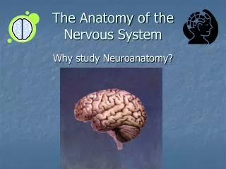
Chapter 4 Anatomy of the Nervous System. Structure of the Vertebrate Nervous System. Neuroanatomy is the anatomy of the nervous system. Refers to the study of the various parts of the nervous system and their respective function(s).

E N D
Structure of the Vertebrate Nervous System • Neuroanatomy is the anatomy of the nervous system. • Refers to the study of the various parts of the nervous system and their respective function(s). • The nervous system consists of many substructures, each comprised of many neurons.
Structure of the Vertebrate Nervous System • Terms used to describe location when referring to the nervous system include: • Ventral: toward the stomach • Dorsal: toward the back • Anterior: toward the front end • Posterior: toward the back end • Lateral: toward the side • Medial: toward the midline
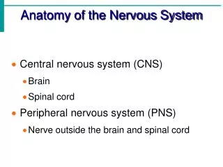
Structure of the Vertebrate Nervous System • The Nervous System is comprised of two major subsystems: • The Central Nervous System (CNS) • The Peripheral Nervous System (PNS)
Structure of the Vertebrate Nervous System • The Central Nervous System consists of: • Brain • Spinal Chord
Structure of the Vertebrate Nervous System • The spinal cord is the part of the CNS found within the spinal column and communicates with the sense organs and muscles below the level of the head. • The Bell-Magendie law states the entering dorsal roots carry sensory information and the exiting ventral roots carry motor information. • The cell bodies of the sensory neurons are located in clusters of neurons outside the spinal cord called dorsal root ganglia.
Structure of the Vertebrate Nervous System • The spinal cord is comprised of: • grey matter-located in the center of the spinal cord and is denseley packed with cell bodies and dendrites • white matter – composed mostly of myelinated axons that carries information from the gray matter to the brain or other areas of the spinal cord. • Each segment sends sensory information to the brain and receives motor commands.
Structure of the Vertebrate Nervous System • The Peripheral Nervous System (PNS) is comprised of the: • Somatic Nervous System • Autonomic Nervous System
Structure of the Vertebrate Nervous System • The Somatic Nervous System consists of nerves that: • Convey sensory information to the CNS. • Transmit messages for motor movement from the CNS to the body.
Structure of the Vertebrate Nervous System • The autonomic nervous system regulates the automatic behaviors of the body (heart rate, blood pressure, respiration, digestion etc). • The autonomic nervous system can be divided into two subsystems: • The Sympathetic Nervous System. • The Parasympathetic Nervous System.
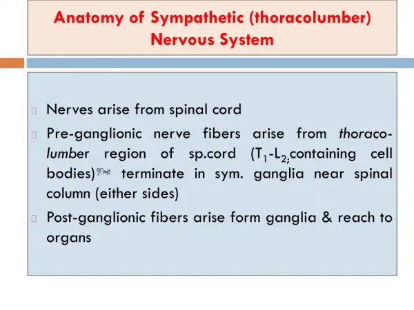
Structure of the Vertebrate Nervous System • The sympathetic nervous system is a network of nerves that prepares the organs for rigorous activity: • increases heart rate, blood pressure, respiration, etc. (“fight or flight” response) • comprised of ganglia on the left and right of the spinal cord • mainly uses norepinephrine as a neurotransmitter at the postganglionic synapses.
Structure of the Vertebrate Nervous System • The parasympathetic nervous system facilitates vegetative, nonemergency responses by the organs. • decreases functions increased by the sympathetic nervous system. • comprised of long preganglion axons extending from the spinal cord and short postganglionic fibers that attach to the organs themselves. • dominant during our relaxed states.
Structure of the Vertebrate Nervous System • Parasympathetic Nervous System (cont’d) • Postganglionic axons mostly release acetylcholine as a neurotransmitter
Structure of the Vertebrate Nervous System • The brain can be divided into three major divisions: • Hindbrain. • Midbrain. • Forebrain.
Structure of the Vertebrate Nervous System • The Hindbrain consists of the: • Medulla. • Pons. • Cerebellum. • Located at the posterior portion of the brain • Hindbrain structures, the midbrain and other central structures of the brain combine and make up the brain stem.
Structure of the Vertebrate Nervous System • The medulla: • Located just above the spinal cord and could be regarded as an enlarged extension of the spinal cord. • responsible for vital reflexes such as breathing, heart rate, vomiting, salivation, coughing and sneezing. • Cranial nerves allow the medulla to control sensations from the head, muscle movements in the head, and many parasympathetic outputs to the organs.
Structure of the Vertebrate Nervous System • Pons • lies on each side of the medulla (ventral and anterior). • along with the medulla, contains the reticular formation and raphe system. • works in conjunction to increase arousal and readiness of other parts of the brain.
Structure of the Vertebrate Nervous System • The reticular formation: • descending portion is one of several brain areas that control the motor areas of the spinal cord. • ascending portion sends output to much of the cerebral cortex, selectively increasing arousal and attention. • The raphe system also sends axons to much of the forebrain, modifying the brain’s readiness to respond to stimuli.
Structure of the Vertebrate Nervous System • The Cerebellum: • a structure located in the hindbrain with many deep folds. • helps regulate motor movement, balance and coordination. • is also important for shifting attention between auditory and visual stimuli.
Structure of the Vertebrate Nervous System • The midbrain is comprised of the following structures: • Tectum – roof of the midbrain • Superior colliculus &inferior colliculus– swellings on each side of the tectum and routes for sensory information • Tagmentum- the intermediate level of the midbrain • Substantia nigra - gives rise to the dopamine-containing pathway
Structure of the Vertebrate Nervous System • The forebrain is the most anterior and prominent part of the mammalian brain and consists of two cerebral hemispheres • Consists of the outer cortex and subcortical regions. • outer portion is known as the “cerebral cortex”. • Receives sensory information and controls motor movement from the opposite (contralateral) side of the body.
Structure of the Vertebrate Nervous System • Subcortical regions are structures of the brain that lie underneath the cortex. • Subcortical structures of the forebrain include: • Thalamus - relay station from the sensory organs and main source of input to the cortex. • Basal Ganglia - important for certain aspects of movement.
Structure of the Vertebrate Nervous System • The limbic system consists of a number of other interlinked structures that form a border around the brainstem. • Includes the olfactory bulb, hypothalamus, hippocampus, amygdala, and cingulate gyrus of the cerebral cortex • associated with motivation, emotion, drives and aggression.
Structure of the Vertebrate Nervous System • Hypothalamus • Small area near the base of the brain. • Conveys messages to the pituitary gland to trigger the release of hormones. • Associated with behaviors such as eating, drinking, sexual behavior and other motivated behaviors. • Thalamus and the hypothalamus together form the “diencephalon”.
Structure of the Vertebrate Nervous System • Pituitary gland - hormone producing gland found at the base of the hypothalamus. • Basal Ganglia - comprised of the caudate nucleus, the putamen and the globus pallidus. • Associated with planning of motor movement, and aspects of memory and emotional expression .
Structure of the Vertebrate Nervous System • Basal forebrain is comprised of several structures that lie on the dorsal surface of the forebrain. • Contains the nucleus basalis: • receives input from the hypothalamus and basal ganglia • sends axons that release acetylcholine to the cerebral cortex • Key part of the brains system for arousal, wakefulness, and attention
Structure of the Vertebrate Nervous System • Hippocampus is a large structure located between the thalamus and cerebral cortex. • critical for storing certain types of memory.
Structure of the Vertebrate Nervous System • The central canal is a fluid-filled channel in the center of the spinal cord. • The ventricles are four fluid-filled cavities within the brain containing cerebrospinal fluid. • Cerebrospinal fluid is a clear fluid similar to blood plasma found in the brain and spinal cord: • Provides “cushioning” for the brain. • Reservoir of hormones and nutrition for the brain and spinal cord.
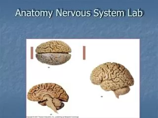
The Cerebral Cortex • The cerebral cortex is the most prominent part of the mammalian brain and consists of the cellular layers on the outer surface of the brain. • comprised of grey matter and white matter. • divided into two halves • joined by two budndles of axons called the corpus callosum and the anterior commissure. • more highly developed in humans than other species.
The Cerebral Cortex • Organization of the Cerebral Cortex: • Contains up to six distinct laminae(layers) that are parallel to the surface of the cortex. • Cells of the cortex are also divided into columnsthat lie perpendicular to the laminae. • Divided into four lobes: occipital, parietal, temporal, and frontal.