Download
1 / 28
280 likes | 385 Views
This study focuses on improving lesion segmentation in brain imaging data using advanced filtering and editing techniques. The process involves manual editing of z-score masks, followed by further refinement and thresholding to create accurate lesion maps, enhancing the quality of the overall analysis. The workflow and outcomes of the editing process are detailed, highlighting the challenges and advancements in lesion boundary identification.

E N D
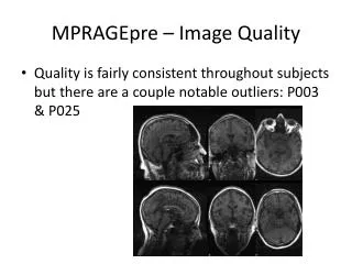
MPRAGEpre – Image Quality • Quality is fairly consistent throughout subjects but there are a couple notable outliers: P003 & P025
Process • Z-score-based (> 4) one-sided thresholding method on FLAIR registered to MNI 1mm3 • > “raw MNI FLAIR z-score masks” • Similar to tissue segmentation, this seemed to contain speckling that was not clearly associated with lesions to me • Median filtering • > “filtered MNI FLAIR z-score masks” • Used median filtering with a kernel of (3mm)3 to reduce speckle • I presented Hagen with the choice of editing either the raw filtered masks
Process • Manual editing • Hagen decided to go work from the raw MNI FLAIR z-score masks • He found the median filtering to be overly aggressive and saw what I had considered as speckling as legitimate lesions • Basic workflow was two passes: remove non-lesion matter such as misclassified skull, add back and correct any lesion boundaries • Hagen focused heavily on the first step and after consideration, felt that most of the pre-lesion boundaries were adequate and actually advantageous because they were not generated by subjective human eyes
Post-Process • Warping back to patient space, SPGR target • Thresholding to recover a binary lesion mask • Further editing?
Quality of Edits • Free of any obvious skull defects • Many lesions remain on sulci, suspicious given limits of standard space registration
Thresholding • Relapsing case, P001: • Some lesions are very close to 0 in value, hard to tell if they should be thrown out • Progressive case, P021: • Thresholds of 0.4-0.6 preserve the sometimes smooth transition into surrounding normal tissue • For conservative lesion boundaries, 0.95 or higher • After we settle on a value, compute the volume of “lost” voxels as a measure of how significant the reduction was
Remaining Work • Tissue segmentation correction • Reiss group multi-spectral segmentation as an alternative to our lesion masks