Download
1 / 28
280 likes | 355 Views
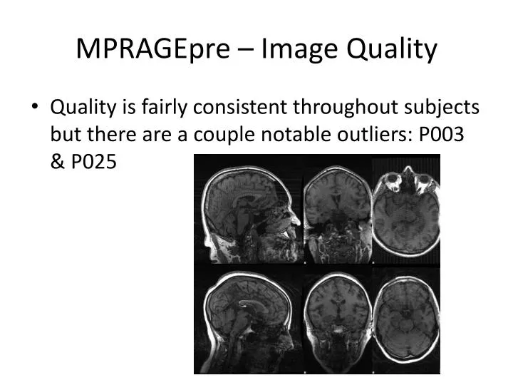
MPRAGEpre – Image Quality. Quality is fairly consistent throughout subjects but there are a couple notable outliers: P003 & P025. Lesion Segmentation. Process. Z-score-based (> 4) one-sided thresholding method on FLAIR registered to MNI 1mm 3 > “raw MNI FLAIR z -score masks”

E N D
MPRAGEpre – Image Quality • Quality is fairly consistent throughout subjects but there are a couple notable outliers: P003 & P025
Process • Z-score-based (> 4) one-sided thresholding method on FLAIR registered to MNI 1mm3 • > “raw MNI FLAIR z-score masks” • Similar to tissue segmentation, this seemed to contain speckling that was not clearly associated with lesions to me • Median filtering • > “filtered MNI FLAIR z-score masks” • Used median filtering with a kernel of (3mm)3 to reduce speckle • I presented Hagen with the choice of editing either the raw filtered masks
Process • Manual editing • Hagen decided to go work from the raw MNI FLAIR z-score masks • He found the median filtering to be overly aggressive and saw what I had considered as speckling as legitimate lesions • Basic workflow was two passes: remove non-lesion matter such as misclassified skull, add back and correct any lesion boundaries • Hagen focused heavily on the first step and after consideration, felt that most of the pre-lesion boundaries were adequate and actually advantageous because they were not generated by subjective human eyes
Post-Process • Warping back to patient space, SPGR target • Thresholding to recover a binary lesion mask • Further editing?
Quality of Edits • Free of any obvious skull defects • Many lesions remain on sulci, suspicious given limits of standard space registration
Thresholding • Relapsing case, P001: • Some lesions are very close to 0 in value, hard to tell if they should be thrown out • Progressive case, P021: • Thresholds of 0.4-0.6 preserve the sometimes smooth transition into surrounding normal tissue • For conservative lesion boundaries, 0.95 or higher • After we settle on a value, compute the volume of “lost” voxels as a measure of how significant the reduction was
Remaining Work • Tissue segmentation correction • Reiss group multi-spectral segmentation as an alternative to our lesion masks