Peripheral angioplasty Overview, Hardware
1.6k likes | 2.33k Views
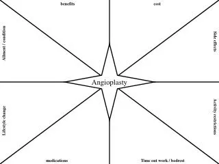
Peripheral angioplasty Overview, Hardware. Frijo Jose A. Vascular Access. Relatively disease-free, without signi Ca Over a bony structure, if possible Angle of entry- 30⁰-45⁰ If access vessel-small/potentially diseased- micropuncture tech preferred. Vascular Access sites.

Peripheral angioplasty Overview, Hardware
E N D
Presentation Transcript
Peripheral angioplastyOverview, Hardware Frijo Jose A
Vascular Access • Relatively disease-free, without signi Ca • Over a bony structure, if possible • Angle of entry- 30⁰-45⁰ • If access vessel-small/potentially diseased- micropuncture tech preferred
Vascular Access sites Retrograde Common Femoral Artery Access • Common access site used for peripheral diagnostic angiography and intervention • Prevent injury to the less diseased extremity
Vascular access sites • Contralateral femoral retrograde access : • Internal iliac stenoses are best treated from a contralateral approach • SFA,PFA- lesions located within the CFA/involve SFA/PFA ostium - • Proximity to arterial puncture site, Bifurcation anatomy of CFA • Also allows treatment B/L disease with a single arterial puncture
Vascular Access site Antegrade Common Femoral Artery Access: • Required for infrainguinal proced • Approx 3cm CFA lies betw ligament & FA bifurcation • Inorder to access CFA, skin entry- prox to ing ligm • Access too close to F bifurc –inadeq working room to selectively cath SFA
Vascular access sites • Ipsilateral popliteal retrograde access: • Useful in SFA occlusion with failure to cross from contralateral or antegrade • Ostial SFA/CFA lesions may also be approached via PA in acute angled terminal aobifurc • CI- aneurysms of PA, pathology of poplitealfossa- Baker’s cyst
Brachial Artery Access • Pref access for visc arterial [CA, SMA] interventions • PC approach at BA can lead to a ↑compli rate • UL arts – smaller, prone to spasm • A small hematoma- Could lead to brachial plexopathy • Itvreq >6F sheaths/smaller pt→open approach preferred • Left BA access pref over Rt- can avoid carotid origin • A micropuncture tech should be used for all PC BA intervention
Wire selection • Many-Teflon/silicone :Some- hydrophilic • Hydr-stenosd/torturous+angle tip–Glidewire • Can be used for crossing tight lesions and can be advanced independent of a guidewire • 014,018,025,035,038-for initial access, 038:18g needle, 018:21g needle
Guidewire-Lesion Interaction • Floppy portion moving in a linear • Floppy portion piles up prox to lesion—no chance to cross- backup,redirect,if straight tip→steerable • Floppy tip bent with min R—Cautiously adv wire- once crossed, wire should straighten- advancing a “buckledup” wire- force→embolization • Floppy tip “buckledup” with R—backup,redirect,adv -dissect,embolz,wiredamag
Catheter ( diagnostic/ guiding) Length depends on location for using a) abdominal aorta = 60 to 80 cm length b) BTK,carotid or subclavian areas 100 to 125cm length Polyethylene- ↓coef friction, pliable Polyurethane- softer, even ↑pliable→ tracks wires better Nylon- stiffer, can tolerate ↑flow rate- amenable to angio Teflon- stiffest- used mainly for dilators & sheaths
▫ ▫
▫ ▫ ▫ ▫ ▫ ▫
4F IMPRESS Simmons 1 Catheter 65cm..038 • Side Ports:N/A • Catheter Shape:SIMMONS1 • French Size:4
5F IMPRESS Simmons 2 Catheter 65cm..038 • Side Ports:N/A • Catheter Shape:SIMMONS2 • French Size: 5
SOS Omni selective catheter • Soft, atraumatic, Super-radiopaque tip • Reforming in descthoracic aorta – below great vessels rather than transverse arch –safety • The catheter should be pulled from the descaointo abdaowith a floppy guidewire“leading,” sometimes with a rotating motion • The soft, flexible atraumatic tip can be placed deeper into the artery (>1 cm), ↓chance of “catheter kickout.” • The shaped tip allows the guidewire to flick into the origin of the RA
Omni Flush Angiographic Catheter • Designed as a single catheter to perform flush aortography, B/L“runoff” studies of lower extremities and to cross aobifurcation with ease for C/L diagnostics in interventional procedures. • Super-Radiopaque tip • Reforms and maintains shape—even under injection pressure—with less catheter whipping, resulting in less vessel wall injury • Less contrast reflux than other flush catheters, thus resulting in lower total contrast dose
Accesses and Selective Guiding Catheters for Some Basic Interventions
Carotid Artery 1.First choice access—either FA 2.Alternative access—left BA 3.Selective catheter— Right carotid: H1,Simmons,Vick; Left carotid : angled glidecath,H1,Simmons Subclavian Artery 1.First choice—either FA 2.Alternative access—ipsilateral BA 3.Selective catheter– angled Glidecath,H1,Simmons,H3 Celiac or SMA 1.First choice—either FA 2.Alternative access—left BA 3.Selective catheter—RIM,Chuang-C,Chuang-3
Renal Artery 1.First choice—contralateral FA 2.Alternative access—left BA 3.Selective catheter—C2,RDC,Sos-omni Infrarenal Aorta 1.First choice —either FA 2.Alternative access—left BA 3.Selective catheter—omni-flush,RIM,C2 Superior Femoral Artery 1.First choice—contralateral FA 2.Alternative—ipsi retro FA for run-off; ipsiantegrade for interv 3.Selective catheter—Berenstein,Kumpe,Vertebral Tibial Arteries 1.First choice—contralateral FA 2.Alternative—ipsi retro FA for run-off; ipsiantegrade for interv 3.Selective catheter—Kumpe,Vertebral
Guiding Catheter vs Sheath • The use of a guide or sheath is determined by operator bias • Sheaths are designed with a simple diaphragm or a hemostatic valve, guiding catheters always require hemostatic valves be attached • During intervention, the guide catheter or sheath should be placed near the lesion to provide for better visualization and improved support
Balloons • In selecting a balloon, the following criteria should be considered: a) Guidewire ( 0.014“, 0.018“, 0.035“) b) over the wire (OTW) or monorail system c) shaft length • 0.014“ balloon system is usually for carotid, vertebral, renal, infrapopliteal arteries • 0.035“ balloon system for subclavian, innominate, aortoiliac, superficial femoral artery • 0.018“ balloon system also in SFA, infrapopliteal, depends on what the operator prefers
Law of Laplace • Circumfer force/tension (T) exerted on wall of an inflatdballn ~P within balln & R (T=P×R) • Balln twice R of a smaller balln- twice wall T for given inflation P→D kept constant, T on wall of ballnwill ↑linearly with ↑inflatn P • Larger ballns -require ↓P than smaller ballnsto generate substantial dilating forces • Larger vessels (Ao) require ↓P to dilate & rupture
Balloon cath with a D matchngoutflow vessel beyond lesion • Balloon length should be > lesion • Balloon centered on lesion & inflated slowly • Inflation maintained for 20s- deflated- reinflated 3 inflations of 20s
Subintimal angioplasty • Hydrophilic wire not passng • Carefully adv into subintimal plane- if not spontaneously, gentle inflation of balloon at edge of the plaque • Wire traversed the lesion subintimaliy • Hydrophilic catheter or other re-entry device passed OTW to guide it back into lumen • Standard angioplasty of subintimal plane performed, with stent placement
Femoropopliteal Artery Intervention Subintimal angioplasty
Stents • The types of stent used in peripheral interventions: • Balloon-expandable • Self-expandable • Stent graft
Balloon-expandable stents • Require positive pressure for expansion • Typically rigid with high radial force • Size of the balloon-expandable stent equals to the size of the reference vessel diameter • Ideal for immobile parts of the body-ie, subclavian, renal, mesenteric, iliac arteries and at ostial locations
Self-expandable Stents • Deployed in vessels that are flexible or twist during movement of neck, shoulder or leg • carotid, axillary, superficial femoral artery, popliteal artery • Nitinol - best flexibility and memory • Stent compressed over a delivery cath & covered with sheath • Stent deployment achieved by pulling back the sheath • Stent diameter should be 1-2mm > ref vessel D→ adeq stent apposition
Self-expandable Stents • Some degree of foreshortening- to be taken into account when choosing • More difficult to place with absolute precision • Generally comes in longer length than BES • Their ability to continually expand after delivery allows them to accommodate adjacent vessels of different size
Stents Demonstrating the Nitinol self-expandable stent deployment
Stent Grafts • Used to exclude aneurysm, treat perforations when prolonged balloon inflation failed • Wallgraft and Viabahn are two options
Decision between SE or BE stents in Iliac Lesions • Balloon expandable • Aortoiliac bifurcation • Common iliac • Calcified lesions • Chronic occlusions (?) • Self expanding • Vessels flexible/twist during movement • Tortuous vessels • Distal external iliac artery • Contralateral approach • Long diffuse lesions • Aortoiliac bifurcation (long lesions)
Techniques Retrograde Iliac stent placement
Techniques Cross-over technique
A patient’s complaint of low back pain during balloon inflation may be a warning sign of adventitial stretch, which may occur before aortic rupture
Femoropopliteal Artery Intervention • Balloon size & length matched to the size ( ~5-6mm) & lesion length( ~40- 300mm) of SFA • ↑ angiographic results may be accomplished with prolonged inflation times ( 3-5 minutes) • Dissections are commonly seen after balloon dilation ( due to heavy calcification)
Femoropopliteal Artery Intervention Stentimplantion ( always SX-Stents): • Sizing the SX- stent ~ 1mm > SFA • Postdilation with 5.0-6.0 mm diameter balloon • Popliteal artery -> avoid stent = high risk of stent compression or fracture
Infrapopliteal Interv • Knee-to-foot patency of one of the three branches is usually sufficient to prevent critical lower-limb ischemia • Claudication is rarely the result of isolated disease of the infrapoplitealarteries • Re-stenosis after intervention in these vessels is typically the highest among the lower limb sites • Obstructive disease in these arteries is often occlusive, diffuse and complicated by heavy calcific deposits
Infrapopliteal Interv- wire selection • Only atraumatic 0.014“ / 0.018“ guide wires should be used-0.014“ prefered due to vessel diameter • Type selection ( floppy, medium,stiff) will be driven by the type of disease
Infrapopliteal -Balloon Angioplasty • Low profile balloon with high pushability and trackability to easy cross the lesion • Flexibility in small collateral branches • 0.014”/ 0.018" wire compatibility • Diameter 1.5mm-4.0mm • Long (20-210 mm) to reduce procedure times and dissection
Infrapopliteal- Balloon Angioplasty Long balloons (210mm/ tapered) • Reducedriskofdissections • ( noballoonoverlap) • Total intervention /revascularization • time significantly shorter • Reduced X-ray dose for patients, operators • as well as for the assistants
Renal artery stenosis • Usually occurs in the proximal 2 cm • ~75% of lesions are caused by atherosclerosis • Lesions can be single or multiple, unilateral or bilateral (~25%) • Diameter: 6.0-6.5mm for men • 5.5-6.0mm for women • Length 3-7 cm
Renal artery-Equipment Diagnostic • Wires • 0.035” for catheter placement • Diagnostic catheter Intervention • Wires • 0.014” • 0.035” for catheter placement • Guiding Sheath • Guide Catheter • Balloons ( 0.014” compatible) • Low profile • Undersized for pre-dilation • BE-Stents