Norepinephrine-induced ROS Production in Cardiomyocytes
Investigating the effect of norepinephrine on ROS production in H9c2 cells and fetal hearts, highlighting the role of NAC, Apo, and DPI as potential mitigators. Data analysis includes comparisons of Egr-1 and Sp1 protein abundance post-treatment. Insights on the expression of Nox1, Nox2, and Nox4 in the context of norepinephrine stimulation.

Norepinephrine-induced ROS Production in Cardiomyocytes
E N D
Presentation Transcript
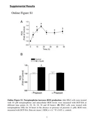
A Supplemental Results Online Figure S1 B Online Figure S1. Norepinephrine increases ROS production. (A): H9c2 cells were treated with 10 M norepinephrine and intracellular ROS levels were measured with DCF-DA at different time points (4, 10, 16, 24, 36 and 48 hours). (B) H9c2 cells were treated with norepinephrine (NE) for 16 hours in the absence or presence of prazosin (1 M). ROS were measured with DCF-DA. Data are mean SEM, n = 6. * P < 0.05 vs. control. 1
Online Figure S2 A B Online Figure S2. Norepinephrine increases ROS production. H9c2 cells and isolated intact fetal hearts (ex vivo) were treated with 10 M norepinephrine (NE) in the absence or presence of N-acetylcysterine (NAC, 1 mM), apocynin (Apo, 0.5 mM) or diphenyleneiodonium (DPI, 10 μM) for 16 hours. ROS were measured with the quantification of confocal images of dihydroethidium (DHE) fluorescence. (A): H9c2 cells. (B): ex vivo fetal hearts. Data are mean SEM, n = 8. * P < 0.05 vs. control. 2
Online Figure S3 Online Figure S3. Effect of norepinephrine on Egr-1 and Sp1 protein abundance. H9c2 cells were treated with 10 M norepinephrine (NE) for 48 hours. Cytosol and nuclear protein abundance of Egr-1 and Sp1 was determined with Western blot. Data are mean SEM, n = 4. 3
H9c2 aorta Protein: 40 60 80 40 g Nox2 Fetalhearts aorta Nox1 Nox2 Nox4 -actin actin Control NE Nox2 actin Online Figure S4 A B C Online Figure S4. Expression of Nox1, Nox2 and Nox4 in fetal hearts and H9c2 cells. (A): Nox2 protein expression was readily detected in rat aortic smooth muscle but not in H9c2 cells. (B): mRNA of Nox1 and Nox4, but not Nox2, was detected in H9c2 cells. (C): Nox2 protein expression was detected in rat aortic smooth muscle but not in isolated fetal rat hearts in the absence or presence of 10 M norepinephrine (NE) for 48 hours. 4