Download
1 / 28
320 likes | 465 Views
The pleural cavity contains fluid that can lead to effusions causing breathing impairments. Learn types, diagnosis, imaging, specimen collection methods, contraindications, and more. Understand transudates vs. exudates, gross and microscopic examination for proper diagnosis.

E N D
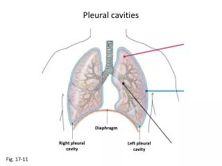
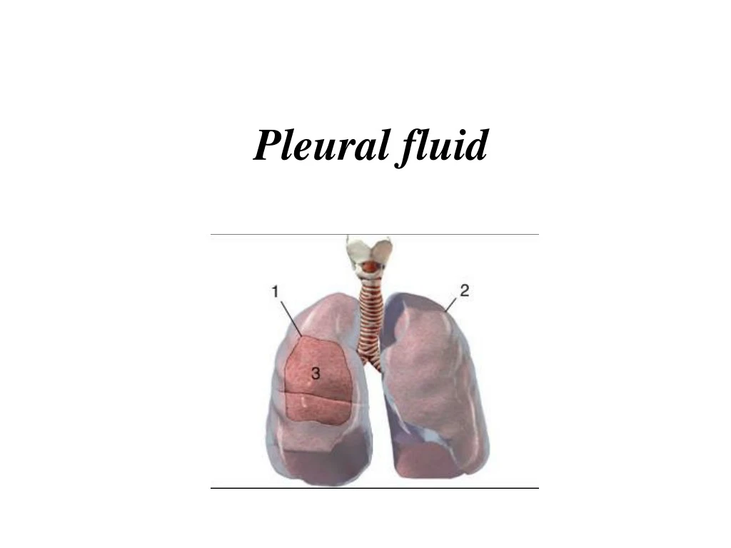
The pleural cavity is a potential space lined by mesothelium of the visceral and parietal pleurae. • The pleural cavity normally contains a small amount of fluid. • This fluid is a plasma filtrate derived from capillaries of the parietal pleura. • It is produced continuously at a rate dependent on capillary hydrostatic • pressure, plasma oncotic pressure, and capillary permeability • Pleural fluid is reabsorbed through the lymphatics and venules of the visceral pleura.
An accumulation of fluid, called an effusion, results from an imbalance of fluid production and reabsorption. • Excessive amounts of such fluid can impair breathing by limiting the expansion of the lungs during ventilation.
Types of fluids • Four types of fluids can accumulate in the pleural space: • Serous fluid (hydrothorax) • Blood(haemothorax ( • Chyle (chylothorax) • Pus (pyothoraxorempyema(
Diagnosis • Pleural effusion is usually diagnosed on the basis ofmedical history andphysical exam, and confirmed bychest x-ray. • Once accumulated fluid is more than 300 ml, there are usually detectableclinical signsin the patient, such as • Decreased movement of the chest on the affected side, • Stony dullness to percussion over the fluid, • Diminishedbreath soundson the affected side, • In large effusion there may betracheal deviation away from the effusion.
Imaging A pleural effusion will show up as an area of whiteness on a standard posteroanterior X-ray. Chest radiographs acquired in the lateraldecubitusposition (with the patient lying on his side) are more sensitive and can pick up as little as 50 ml of fluid. At least 300 ml of fluid must be present before upright chest films can pick up signs of pleural effusion (e.g., bluntedcostophrenic angles)
Massive left sided pleural effusion in a patient presenting with lung cancer.
CT scan of chest showing loculated pleural effusion in left side. Some thickening of pleura is also noted.
SPECIMEN COLLECTION • Thoracentesis is indicated for any undiagnosed pleural effusion or for therapeutic purposes in patients with massive symptomatic effusions; • A needle is inserted through the back of the chest wall in the sixth, seventh, or eighth intercostal space on the midaxillary line, into the pleural space. • The fluid may then be evaluated for the following: • Chemical composition includingprotein, lactat dehydrogenaseLDH, albumin, amylase, pH, andglucose. • Gram stainand culture to identify possible bacterial infections • Cellcount and differential • Cytopathologyto identify cancer cells, but may also identify some infective organisms • Other tests as suggested by the clinical situation – lipids, fungal culture, viral culture, specificimmunoglobulins
Contraindications • An uncooperative patient or acoagulationdisorder that can not be corrected are absolute contraindications • Relative contraindications include cases in which the site of insertion has known bullous disease (e.g. emphysema( and use ofmechanical ventilation.
TRANSUDATES AND EXUDATES Transudates are usually bilateral owing to systemic conditions leading to increased capillary hydrostatic pressure or decreased plasma oncotic pressure Exudates are more often unilateral, associated with localized disorders that increase vascular permeability or interfere with lymphatic resorption
Classical teaching stressed that exudates and transudates can be distinguished on the basis of total protein concentrations above (exudates) or below (transudates) 3.0 g/dL • Accordingly, an exudate meets one or more of the following criteria: • Pleural fluid/serumv protein ratio greater than 0.5; • pleural fluid/serum LD ratio greater than 0.6; and • pleural fluid LD level greater than two thirds of the serum upper limit of normal.
GROSS EXAMINATION Transudates are typically clear, pale yellow to straw-colored, and odorless, and do not clot. Approximately 15% of transudates are blood tinged. A bloody pleural effusion (hematocrit >1%) suggests trauma, malignancy, or pulmonary infarction. A pleural fluid hematocrit greater than 50% of the blood hematocrit is good evidence for a hemothorax
Exudates may grossly resemble transudates, but most show variable degrees of cloudiness or turbidity, and they often clot if not heparinized. A feculent odor may be detected in anaerobic infections. Turbid, milky, and/or bloody specimens should be centrifuged and the supernatant examined. If the supernatant is clear, the turbidity is most likely due to cellular elements or debris. If the turbidity persists after centrifugation, a chylous effusion is likely.
MICROSCOPIC EXAMINATION Cell Counts Leukocyte counts have limited utility in separating transudates (<1000/μL) from exudates (>1000/μL). Although red cell counts above 100,000/μL are highly suggestive of malignancy, trauma, or pulmonary infarction, they are not specific for these conditions.
Differential Leukocyte Count and Cytology Cytologic analysis will establish the diagnosis of metastatic carcinoma in 70% or more of cases
CHEMICAL ANALYSIS Protein The measurement of pleural fluid total protein or albumin has little clinical value except when combined with other parameters to differentiate exudates from transudates. Glucose The glucose level of normal pleural fluid, transudates, and most exudates is similar to serum levels. Decreased pleural fluid glucose, accepted as a level below 60 mg/dL (3.33 mmol/L) or a pleural fluid/serum glucose ratio less than 0.5, is most consistent and dramatic in rheumatoid pleuritis and grossly purulent parapneumonic exudates
Lactate Pleural fluid lactate levels can be a useful adjunct in the rapid diagnosis of infectious pleuritis. Levels are significantly higher in bacterial and tuberculous pleural infections than in other pleural effusions. Values greater than 90 mg/dL (10 mmol/L) have a positive predictive value for infectious pleuritis of 94% and a negative predictive value of 100%. Amylase: elevations above the serum level (usually 1.5–2.0 or more times greater) indicate the presence of pancreatitis, esophageal rupture, or malignant effusion. Elevated amylase derived from esophageal rupture or malignancy is the salivary isoform, which differentiates it from pancreatic amylase
Lactate dehydrogenase Pleural fluid LD levels rise in proportion to the degree of inflammation. In addition to their use in separating exudates from transudates, declining LD levels during the course of an effusion indicate that the inflammatory process is resolving. Conversely, increasing levels indicate a worsening condition requiring aggressive workup or treatment. Interferon-γ: Pleural fluid interferon (IFN)-γ levels are significantly increased in the pleural fluid of patients with tuberculous pleuritis. The sensitivity of levels of 3.7 IU/L or greater is 99%, and the pecificity is 98%
pH Pleural fluid pH measurement has the highest diagnostic accuracy in assessing the prognosis of parapneumonic (pneumonia-related) effusions A parapneumonic exudate with a pH greater than 7.30 generally resolves with medical therapy alone. A pH less than 7.20 indicates a complicated parapneumonic effusion (loculated or associated with empyema), requiring surgical drainage. A pH below 6.0 is characteristic of esophageal rupture, although the pH in severe empyema may be 6.0 or less
Lipids Lipid measurements are also helpful in identifying chylous effusions Thus, pleural fluid triglyceride levels above 110 mg/dL indicate a chylous effusion; values from 60–110 mg/dL (0.68–1.24 mmol/L) are less certain and require lipoprotein electrophoresis to confirm a chylothorax. Nonchylous effusions generally have triglyceride levels below 50 mg/dL (0.56 mmol/L) and no chylomicrons on electrophoresis Cholesterol measurements may be useful in separating transudates from exudates, especially when there is a question regarding Light’s criteria A total cholesterol value of 54 mg/dL or more and a pleural fluid/ serum cholesterol ratio of 0.32 or higher have sensitivity and specificity values similar to Light’s criteria
IMMUNOLOGIC STUDIES Approximately 5% of patients with RA and 50% with SLE develop pleural effusions sometime during the course of their disease. RF is commonly present in pleural effusions associated with seropositive RA. ANA titers may be useful in the diagnosis of effusion due to lupus pleuritis
MICROBIOLOGICAL EXAMINATION Bacteria most commonly associated with parapneumonic effusions are Staphylococcus aureus, Streptococcus pneumoniae, β-hemolytic group A streptococci, enterococci, and some gram-negative bacilli. Anaerobic bacteria are isolated in a significant proportion of cases, so both anaerobic and aerobic cultures should be performed. The sensitivity of the Gram stain is approximately 50% For patients with suspected M. tuberculosis, direct staining of tuberculous effusions for acid-fast bacteria has a sensitivity of 20%–30%, and positive cultures are found in 50%–70% of cases