Download
1 / 14
160 likes | 574 Views
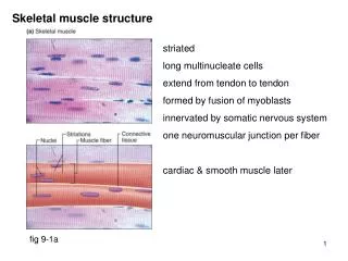
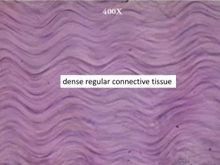
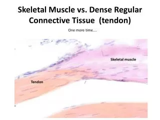
Skeletal Muscle vs. Dense Regular Connective Tissue (tendon). One more time…. Skeletal muscle . Tendon. Muscle Fiber Types . Cross Section of Skeletal M uscle S tained for Myoglobin

E N D
Skeletal Muscle vs. Dense Regular Connective Tissue (tendon) • One more time…. Skeletal muscle Tendon
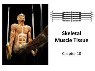
Muscle Fiber Types
Cross Section of Skeletal Muscle Stained for Myoglobin -Type I Fibers (aka. Red Fibers, Slow Oxidative): Darkly staining b/c more myoblobin, slow twitch, fatigue resistant, but generate less tension -Type IIa Fibers (aka. Intermediate fibers, fast glycolytic oxidative fibers): Variable staining, fast-twitch, fatigue resistant, capable of anaerobic glycolysis - Type IIb Fibers (aka. White Fibers, Fast Glycolytic): Lightest staining, fast-twitch, fatigue prone, lots of glycogen, low myoglobin, round, large White Red
Motor End Plate -where black staining nerves contact muscle -looks like the rubber part of a plunger
Peripheral Nerves: -In the accurate words of the legendary Dr. Boggus: “Circle of wavy shit”
Peripheral Nerves: -Endonuriem: around each axon -Perinuriem: around each peripheral nerve -Epinuerium: around each bundle of peripheral nerves
Peripheral Nerves: -Endonuriem (EN): around each axon -Perinuriem (P): around each peripheral nerve -Epinuerium(EPI): around each bundle of peripheral nerves *from demo on blackboard
Peripheral Nerve: Longitudinal Section -Field of bubbly wavy shit -Nodes of Ranvier are the short vertical lines b/w Shwann cells (ie the ties b/w the sausage links)
Myelinated Nerve: -Nuerilemma: surrounds the myelinated cells -Node of Ranvier: the dark line b/w Shwann cells where the nuerilemma appears pinched. Myelin Axon Myelin
Myelinated Nerve: -Myelin sheath is stained black (the small black circles)
Ganglion: -Looks like a bunch of FRIED EGGS -DON’T confuse this with peripheral nerve (circle of W.S.)! In a peripheral nerve cross section, the dark spot is an axon, not a nucleus w/ a nucleolus, and the white of the egg is a pale myelin sheath with a dark outline as opposed to cell cytoplasm.
Ganglion vs. Peripheral Nerve Ganglion Peripheral Nerve -Circle of W.S. -Fried eggs -Dark axons surrounded by pale myelin sheaths with darker outlines -Lots of satellite cells! -Darkish staining cytoplasm in neurons that tends to look shrunken/pull away from surroundings (ie white space) -I spy a nucleolus!