Download

1 / 46
500 likes | 1.02k Views
Longitudinal fissure. Cerebrum. 6. 1. Gyrus. 2. Central sulcus. Sulcus. 5. 3. Cut. Lateral fissure. 4. 1. Anterior. 2. 3. 4. 5. 6. Station 1 – Whole brain. T. Hints: 1. Brain division 2. Raised area on surface of the cerebrum 3. Depression on surface of the cerebrum

E N D
Longitudinal fissure Cerebrum 6 1 Gyrus 2 Central sulcus Sulcus 5 3 Cut Lateral fissure 4 1 Anterior 2 3 4 5 6 Station 1 – Whole brain T Hints: 1. Brain division 2. Raised area on surface of the cerebrum 3. Depression on surface of the cerebrum 4. Separates superior/inferior cerebral lobes 5. Separates anterior/posterior cerebral lobes 6. Separates cerebral hemispheres • Terms in alphabetic order:Central sulcusCerebrumGyrusLateral fissureLongitudinal fissureSulcus H
Parietal lobe 4 Frontal lobe 1 Occipital lobe 3 Temporal lobe 2 1 Anterior 2 3 4 5 6 Station 1 – Whole brain T Hints: 1. Lobe of the cerebrum 2. Lobe of the cerebrum 3. Lobe of the cerebrum 4. Lobe of the cerebrum • Terms in alphabetic order:Frontal lobeOccipital lobeParietal lobeTemporal lobe H
6 – Function of 5? Commissural nerve tract Corpus callosum 5 Anterior Lateral ventricle 4 Insula 3 1 Temporal lobe Frontal lobe 2 2 1 3 4 5 6 Station 2 – Part of right cerebral hemisphere removed T • Terms in alphabetic order:Corpus callosumFrontal lobeInsulaLateral ventricleTemporal lobe Hints: 1. Lobe of cerebrum 2. Lobe of cerebrum 3. Lobe of cerebrum 4. Space 5. White matter 6. Function of #5? H
Anterior Nerve tract Station 2 – Part of right cerebral hemisphere removed For Your Information: During development, the corpus callosum begins in the anterior part of the brain and grows posteriorly. Some of the anterior brain tissue is dragged posteriorly with the growing corpus callosum. In the model, this tissue is represented by the green stripes on the corpus callosum. In the adult brain, this tissue connects the anterior brain to other parts of the brain. The rest of this tissue will be seen as we “dissect” the brain further.
Anterior Insula 1 Internal capsule 2 3 – Function of 2? Projection nerve tract 1 Lateral ventricle 4 2 3 Corpus callosum 5 4 5 6 Station 2 - Part of right cerebral hemisphere removed T • Terms in alphabetic order:Corpus callosumInsulaInternal capsuleLateral ventricle Hints: 1. Lobe of cerebrum 2. White matter 3. Function of #2? 4. Space 5. White matter H
Central sulcus 1 Premotor area 5 Prefrontal area Primary motor cortex 4 2 Broca’s area 3 1 Anterior 2 3 4 5 6 Station 3 – Functional regions of the left cerebral cortex T • Terms in alphabetic order:Broca’s areaCentral sulcusPrefrontal areaPremotor areaPrimary motor cortex Hints: 1. Separates frontal and parietal lobes 2. Functional region 3. Functional region 4. Functional region 5. Functional region H
Central sulcus 1 Primary somatic sensory cortex 2 Somatic sensory association area 3 Taste area 4 1 Anterior 2 3 4 5 6 Station 3 – Functional regions of the left cerebral cortex T • Terms in alphabetic order:Central sulcusPrimary somatic sensory cortexSomatic sensory association areaTaste area Hints: 1. Depression 2. Functional region 3. Functional region 4. Functional region H
Anterior Auditory association area 4 Visual cortex 1 Visual association area 2 1 Primary auditory cortex 3 2 3 4 5 6 Station 3 – Functional regions of the left cerebral cortex T • Terms in alphabetic order:Auditory association areaPrimary auditory cortexVisual association areaVisual cortex Hints: 1. Functional region 2. Functional region 3. Functional region 4. Functional region H
Primary motor cortex 1 Premotor area 2 Prefrontal area 3 1 2 Anterior 3 4 5 6 Station 3 – Functional regions of the left cerebral cortex T • Terms in alphabetic order:Prefrontal areaPremotor areaPrimary motor cortex Hints: 1. Functional region 2. Functional region 3. Functional region H
Somatic sensory association area Primary somatic sensory cortex 2 1 Visual association area 3 Visual cortex 4 1 2 Anterior 3 4 5 6 Station 3 – Functional regions of the left cerebral cortex T • Terms in alphabetic order:Primary somatic sensory cortexSomatic sensory association areaVisual association areaVisual cortex Hints: 1. Functional region 2. Functional region 3. Functional region 4. Functional region H
Limbic system (part of) 1 Corpus callosum 2 Fornix 3 1 4 – Function of 3? Association nerve tract 2 Temporal lobe 5 3 Anterior 4 5 6 Station 3 – Functional regions of the left cerebral cortex T • Terms in alphabetic order:Corpus callosumFornixLimbic system (part of)Temporal lobe Hints: 1. Functional region 2. White matter 3. White matter 4. Function of #3 5. Lobe of cerebrum H
Limbic system (part of) 1 Corpus callosum (cut) Fornix 2 Choroid plexus 6 Limbic system (part of) 3 Hippocampus 5 1 2 Temporal lobe 4 3 4 Anterior 5 6 Station 4 – Superior part of cerebrum removed T • Terms in alphabetic order:Choroid plexusFornixHippocampusLimbic system (part of)Temporal lobe Hints: 1. Functional region 2. White matter 3. Functional region 4. Lobe of cerebrum 5. Gyrus 6. Specific structure H
Nerve tract Dentate gyrus Anterior Station 4 – Superior part of cerebrum removed For Your Information: The nerve tract passes inferiorly and joins the dentate gryrus (dark green), which is part of the hippocampus (light green).
Anterior Frontal lobe 1 Temporal lobe 2 Location of diencephalon and brainstem Fornix 3 Nerve tract 1 2 3 4 5 6 Station 5 – Inferior view of cerebrum T • Terms in alphabetic order:FornixFrontal lobeTemporal lobe Hints: 1. Lobe of cerebrum 2. Lobe of cerebrum 3. White matter H
Anterior Olfactory bulb 1 Olfactory tract 2 Model peg Primary olfactory cortex 3 (dark brown) 1 Olfactory association area 2 4 3 4 5 6 Station 5 – Inferior view of cerebrum T • Terms in alphabetic order:Olfactory association areaOlfactory bulbOlfactory tractPrimary olfactory cortex Hints: 1. White matter 2. White matter 3. Functional region 4. Functional region H
Anterior Sulcus 6 Gyrus Frontal lobe 5 1 Insula 2 Temporal lobe 3 1 2 Occipital lobe 3 4 4 5 6 Station 6 – Superior part of cerebrum removed T • Terms in alphabetic order:Frontal lobeGyrusInsulaOccipital lobeSulcusTemporal lobe Hints: 1. Lobe of cerebrum 2. Lobe of cerebrum 3. Lobe of cerebrum 4. Lobe of cerebrum 5. Raised area 6. Depression H
Anterior Internal capsule 1 Lateral ventricle 2 Cerebral medulla 5 1 Corpus callosum 3 Cerebral cortex 4 2 3 4 5 6 Station 6 – Superior part of cerebrum removed T • Terms in alphabetic order:Cerebral cortexCerebral medullaCorpus callosumInternal capsuleLateral ventricle Hints: 1. White matter 2. Space 3. White matter 4. Gray matter 5. White matter H
Anterior Internal capsule 1 Corpus callosum (cut) Caudate nucleus 2 Fornix 4 Thalamus 3 Corpus callosum (cut) 1 2 3 4 5 6 Station 7 – Corpus callosum and ventricles removed T • Terms in alphabetic order:Caudate nucleusFornixInternal capsuleThalamus Hints: 1. White matter 2. Gray matter 3. Brain division 4. White matter H
Corpus callosum 1 Anterior 1 2 3 Septa pellucida 2 4 5 6 Station 7 – Lateral view of corpus callosum T • Terms in alphabetic order:Corpus callosumSepta pellucida Hints: 1. White matter 2. Specific structure H
Anterior Internal capsule 6 Thalamus 1 Insula 5 Pineal body 2 Corpora quadrigemina 3 1 2 Cerebellum 4 3 4 5 6 Station 8 – Most of cerebrum removed T • Terms in alphabetic order:CerebellumCorpora quadrigeminaInsulaInternal capsulePineal bodyThalamus Hints: 1. Brain division 2. Specific structure 3. Specific structure 4. Brain division 5. Lobe of cerebrum 6. White matter H
Anterior Folium 1 Fissure 2 1 2 3 Vermis Lateral hemisphere 3 4 4 5 6 Station 8 – Superior view of the cerebellum T • Terms in alphabetic order:Folium (gyrus)Lateral hemisphereSulcus (fissure)Vermis Hints: 1. Raised area 2. Depression 3. One of three parts of the cerebellum 4. One of three parts of the cerebellum H
Arbor vitae 1 Folium (gyrus) 2 Anterior Cortex 3 1 2 3 4 5 6 Station 8 – Median view of cerebellum T • Terms in alphabetic order:Arbor vitaeCortexFolium (gyrus) Hints: 1. White matter 2. Raised area 3. Gray matter H
Cerebellar peduncles 1 Superior 2 – Function of 1? Connect cerebellum to brainstem 1 2 3 Flocculonodular lobe 3 4 5 6 Station 8 – Anterior view of cerebellum T • Terms in alphabetic order:Cerebellar pedunclesFlocculonodular lobe Hints: 1. White matter 2. Function of #1 3. One of three parts of the cerebellum H
Superior sagittal sinus 6 Corpus callosum 1 Dura mater 5 Lateral ventricle Anterior 4 1 2 Cerebellum 3 3 Insula 2 4 5 6 Station 9 - Most of the cerebrum is removed T Hints: 1. White matter 2. Lobe of the cerebrum 3. Brain division 4. Space 5. Membrane 6. Blood vessel • Terms in alphabetic order:CerebellumCorpus callosumDura materInsulaLateral ventricleSuperior sagittal sinus H
Corpus callosum 1 Septa pellucida 2 Thalamus 6 Fornix 3 Cerebellum 5 Brainstem 4 1 2 Anterior 3 4 5 6 Station 10 – Median view of the brain T • Terms in alphabetic order:BrainstemCerebellumCorpus callosumFornixSepta pellucidaThalamus Hints: 1. White matter 2. Partition 3. White matter 4. General area 5. Brain division 6. Brain division H
Location of interthalamic adhesion 2 Third ventricle Lateral ventricle 3 1 Anterior Cerebral aqueduct 4 1 Fourth ventricle 5 2 3 Central canal 6 4 5 6 Station 11 - Ventricles T • Terms in alphabetic order:Central canalCerebral aqueductFourth ventricleLateral ventricleLocation of interthalamic adhesionThird ventricle Hints: 1. Space 2. Location of connection between thalamic lobes 3. Space 4. Space 5. Space 6. Space H
Interthalamic adhesion 2 Lateral ventricle 1 Third ventricle 3 Anterior Cerebral aqueduct 4 1 Fourth ventricle 5 2 3 Central canal 4 6 5 6 Station 11 - Ventricles T • Terms in alphabetic order:Central canalCerebral aqueductFourth ventricleLateral ventricleLocation of interthalamic adhesionThird ventricle Hints: 1. Space 2. Specific structure 3. Space 4. Space 5. Space 6. Space H
Internal capsule Insula Anterior Station 12 – A reminder of the location of the internal capsule and insula
Internal capsule 1 Lentiform nucleus 2 Corpus striatum 4 Caudate nucleus 3 1 2 3 4 Anterior 5 6 Station 12 – Insula removed on left and right sides T • Terms in alphabetic order:Caudate nucleusCorpus striatumInternal capsuleLentiform nucleus Hints: 1. White matter 2. Gray matter 3. Gray matter 4. Number 2 and 3 combined H
Internal capsule 1 Lentiform nucleus 2 Corpus striatum 4 Caudate nucleus 3 1 2 3 4 Anterior 5 6 Station 12 – Most of internal capsule removed on the left T • Terms in alphabetic order:Caudate nucleusCorpus striatumInternal capsuleLentiform nucleus Hints: 1. White matter 2. Gray matter 3. Gray matter 4. Number 2 and 3 combined H
Internal capsule 1 Thalamus 4 Insula 3 Caudate nucleus 2 1 2 3 4 Anterior 5 6 Station 13 – Superior view of central core of the brain T • Terms in alphabetic order:Caudate nucleusInsulaInternal capsuleThalamus Hints: 1. White matter 2. Gray matter 3. Lobe of cerebrum 4. Brain division H
Produces cerebrospinal fluid 3 – Function of 2? Fornix 1 Choroid plexus 2 1 2 3 4 Anterior 5 6 Station 13 – Superior view of central core of the brain T • Terms in alphabetic order:Choroid plexusFornix Hints: 1. White matter 2. Specific structure 3. Function of #2? H
Thalamus 5 Fornix 1 Third ventricle 4 Amygdala 3 1 Optic nerve 2 2 3 4 5 6 Station 13 – Anterosuperior view of central core of the brain T • Terms in alphabetic order:AmygdalaFornixOptic nerveThalamusThird ventricle Hints: 1. White matter 2. White matter 3. Gray matter 4. Space 5. Brain division H
Optic nerve 1 Amygdala 2 Hippocampus 3 Brainstem 4 1 2 3 4 5 6 Station 13 – Anteroinferior view of central core of the brain T • Terms in alphabetic order:AmygdalaBrainstemHippocampusOptic nerve Hints: 1. White matter 2. Gray matter 3. Gyrus of the cerebrum 4. General area H
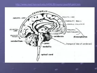
Infundibulum 1 Mammillary body 2 Cerebral peduncle 3 Pons 4 Medulla oblongata 5 1 2 3 4 5 6 Station 14 – Anteroinferior view of central core of the brain T • Terms in alphabetic order:Cerebral peduncleInfundibulumMammillary bodyMedulla oblongataPons Hints: 1. Specific structure 2. Specific structure 3. Specific structure 4. Brain division 5. Brain division H
Pyramid 1 1 Pyramidal decussation 2 2 Spinal cord 3 3 4 5 6 Station 14 – Anteroinferior view of central core of the brain T • Terms in alphabetic order:PyramidPyramidal decussationSpinal cord Hints: 1. Specific structure 2. Specific structure 3. Specific structure H
Fornix 1 Hippocampus 2 Brainstem 3 1 2 3 4 5 6 Station 15 – Posterior view of central core of the brain T • Terms in alphabetic order:BrainstemFornixHippocampus Hints: 1. White matter 2. Gyrus of cerebrum 3. General area H
Pineal body 1 Superior colliculus 2 Corpora quadrigemina 4 Inferior colliculus 3 Midbrain 6 Tegmentum 5 1 2 3 4 5 6 Station 15 – Posterior view of central core of the brain T • Terms in alphabetic order:Corpora quadrigeminaInferior colliculusMidbrainPineal bodySuperior colliculusTegmentum Hints: 1. Specific structure 2. Specific structure 3. Specific structure 4. Number 2 and 3 combined 5. Specific structure 6. Brain division H
Cerebral aqueduct 4 – connects to ? Cerebral peduncles 1 1 Central canal of spinal cord 3 – connects to ? 2 Fourth ventricle 2 3 4 5 6 Station 15 – Posterior view of central core of the brain T • Terms in alphabetic order:Central canal of spinal cordCerebral aqueductCerebral pedunclesFourth ventricle Hints: 1. White matter 2. Space 3. Space 4. Space H
Optic nerve (CN II) 1 Optic chiasm 2 Optic tract 3 Oculomotor nerve (CN III) 4 Trochlear nerve (CN IV) 5 1 2 Trigeminal nerve (CN V) 6 3 4 5 6 Station 16 – Anteroinferior view of central core of the brain T • Terms in alphabetic order:Oculomotor nerve (CNIII)Optic chiasmOptic nerve (CN II)Optic tractTrigeminal nerve (CN V)Trochlear nerve (CN IV) Hints: 1. Cranial nerve 2. White matter 3. White matter 4. Cranial nerve 5. Cranial nerve 6. Cranial nerve H
Trochlear nerve (CN IV) 1 Trigeminal nerve (CN V) 2 Pons 3 Olive 4 1 Anterior 2 3 4 5 6 Station 16 – Lateral view of the brainstem T • Terms in alphabetic order:OlivePonsTrigeminal nerve (CN V)Trochlear nerve (CN (IV) Hints: 1. Cranial nerve 2. Cranial nerve 3. Brain division 4. Specific structure H
Abducent nerve (CN VI) 1 Facial nerve (CN VII) 2 1 2 Vestibulocochlear nerve (CN VIII) 3 3 4 5 6 Station 17 – Anteroinferior view of central core of the brain T • Terms in alphabetic order:Abducent nerve (CN VI)Facial nerve (CN VII)Vestibulocochlear nerve (CN VIII) Hints: 1. Cranial nerve 2. Cranial nerve 3. Cranial nerve H
Vestibulocochlear nerve (CN VIII) 2 Facial nerve (CN VII) 1 Glossopharyngeal nerve (CN IX) 3 Anterior Vagus nerve (CN X) 4 1 2 Hypoglossal nerve (CN XII) Accessory nerve (CN XI) 6 5 3 4 5 6 Station 17 – Lateral view of the brainstem T • Terms in alphabetic order:Accessory nerve (CN XI)Facial nerve (CN VII)Glossopharyngeal nerve (CN IX)Hypoglossal nerve (CN XII)Vagus nerve (CN X)Vestibulocochlear nerve (CN VIII) Hints: 1. Cranial nerve 2. Cranial nerve 3. Cranial nerve 4. Cranial nerve 5. Cranial nerve 6. Cranial nerve H
Anterior Caudate nucleus 1 Nerve tract Interthalamic adhesion 6 Pineal body 2 Superior colliculus 3 Thalamus 5 1 Inferior colliculus 4 2 3 4 5 6 Station 18 – Sagittal section of brainstem T • Terms in alphabetic order:Caudate nucleusInferior colliculusInterthalamic adhesionPineal bodySuperior colliculusThalamus Hints: 1. Gray matter 2. Specific structure 3. Specific structure 4. Specific structure 5. Brain division 6. Specific structure H
Anterior Hypothalamus 6 Fornix 1 Optic chiasm 5 Mammillary body 2 Pons 4 1 Medulla oblongata 3 2 3 4 5 6 Station 19 – Sagittal section of brainstem T • Terms in alphabetic order:FornixHypothalamusMammillary bodyMedulla oblongataOptic chiasmPons Hints: 1. White matter 2. Specific structure 3. Brain division 4. Brain division 5. White matter 6. Brain division H
Anterior Third ventricle 1 Cerebral aqueduct 2 Fourth ventricle 3 1 2 Central canal 4 3 4 5 6 Station 19 – Sagittal section of brainstem T • Terms in alphabetic order:Central canalCerebral aqueductFourth ventricleThird ventricle Hints: 1. Space 2. Space 3. Space 4. Space H