GASTROINTESTINAL SYSTEM PROTOZOA -I-
710 likes | 1.75k Views
GASTROINTESTINAL SYSTEM PROTOZOA -I-. Entamoeba histolytica Giardia lamblia Entamoeba coli. Doç.Dr.Hrisi BAHAR. Gastrointestinal system protozoon. Entamoeba histolytica. Pathogenic Entamoeba histolytica Balantidium coli Giardia lamblia Dientamoeba fragilis Cryptosporidium parvum

GASTROINTESTINAL SYSTEM PROTOZOA -I-
E N D
Presentation Transcript
GASTROINTESTINAL SYSTEM PROTOZOA -I- Entamoeba histolytica Giardia lamblia Entamoeba coli Doç.Dr.Hrisi BAHAR
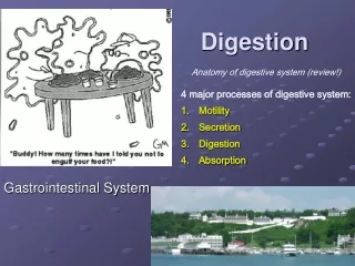
Gastrointestinal system protozoon Entamoeba histolytica
Pathogenic Entamoeba histolytica Balantidium coli Giardia lamblia Dientamoeba fragilis Cryptosporidium parvum Enterocytozoon bieneusi Septata intestinalis Cyclospora cayetanensis Isospora belli Commensal Entamoeba hartmani Entamoeba dispar Entamoeba coli Endolimax nana Iodamoeba bütschlii Chilomastix mesnili Trichomonas hominis Blastocystis hominis INTESTINAL PROTOZOA
Entamoeba histolytica(amoebiasis) ►Organism:Entamoebahistolytica ► At Risk : Anyone worldwide ► Humans Infected: 50 million cases of invasive disease/year ►Disease Outcome :100,000 deaths/year ►Available Drugs :Dose limiting side effects .
Entamoeba histolytica(amoebiasis) Morphologic forms: 1- Active trophozoit form *Cytoplasm:(2/3endoplasm and 1/3 ectoplasm) * Nucleus: circle with karyosome * Replication stage 2-4 nucleus
Entamoeba histolytica(amoebiasis) Active trophozoit form
Entamoeba histolytica(amoebiasis) 2-Precystic form: *Intermediate stage *Low motility *Untrue morphologic stage of parasites *shape: oval or circle
Entamoeba histolytica(amoebiasis) Precystic form
Entamoeba histolytica(amoebiasis) 3-Cystic form: *Stable and non motile *Small and large cysts *Primary cyst with -1- nucleus *In replication stage 2-4 nucleus *Karyosome,, chromatoide body and glycogen in nucleus
Entamoeba histolytica(amoebiasis) Cystic form
Entamoeba histolytica(amoebiasis) Replication stage of cyst
Entamoeba histolytica(amoebiasis) Stability *Trophozoit form: unstable *Cystic form: stablein 20ºC for 3 days and In 45 ºC for 30 min *Cystic form also stable againstlow concentration ofCLandHCL
Entamoeba histolytica(amoebiasis) Transmission *It is transmitted by cystic form* ☻ Direct transmission With contaminated hands ☻ Indirect transmision With contaminated food , water and arthropods.
Entamoeba histolytica(amoebiasis) Pathogenesis ► Cases acute dysentery ► Chronic stage: occur intestinal ulcer,inflammation and necrosis with proteolytic enzyme ► Heavy infection: occur intestine scleroses ,hypertrophy and perforation ► Metastases: to liver and brain…
Entamoeba histolytica(amoebiasis) Intestinal ameobiasis ●Incubation period is 8 days or several month ● Acute stage: diarrhea with epithelium but without blood, and abdominal pine , loss of weight , flatulence and constipation. ● Sever infection : 10-20/day , diarrhea with blood , abdominal pine (colon)dehydration and fever.
Entamoeba histolytica(amoebiasis) Extra intestinal amoebiais ►Spread of intestinal amoeba from blood to liver , spleen,brain and lung. ►Direct extra intestinal amoebiasis (without intestinal infection)
Entamoeba histolytica(amoebiasis) Hepatit Amoebiasis ►Causes liver abscess ►Single abscess, different size , at the right lob of the liver.
Entamoeba histolytica(amoebiasis) Hepatic Amoebiasis ►Symptoms: liver pine , fever right hypochondrium pain,rarely diarrhea ►Transmit to pleura, peritone and pericardial area…..dead.
Entamoeba histolytica(amoebiasis) Hepatic Amoebiasis AMOEBIC LIVER ABSCESS Diagnosis *Ultrasound *Raised WBC *Serology *Aspirate microscopy *Response to metronidazole 750 t.i.d.
Entamoeba histolytica(amoebiasis) Pulmonary Amoebiasis ► 1- Direct primary infection(blood circulation) ► 2- secondary infection: after liver amoebiasis at the right pulmonary .. Cases are with: single or several abscess
Entamoeba histolytica(amoebiasis) Cerebral Amoebiasis ► Occur from complication of liver and pulmonary amoebiasis.. ► Cases are with single or several abscess
Entamoeba histolytica(amoebiasis Spleen and Cutaneous Amoebiasis ►Spleen abscess always seem with liver amoebiasis. ► Cutaneous amoebiasis seem in perianal site.
Entamoeba histolytica(amoebiasis Diagnosis ►Detection of trophozoit and cystic form of parasites in fresh stool. ► After 30min trophozoit form will destroy. ► Extra intestinal amoebiasis: detection of parasite cysts by lugol stain of infected tissue.
Entamoeba histolytica(amoebiasis trophozoit Direct microscopy : Detection of trophozoit and cystic form of parasites in fresh stool.
Entamoeba histolytica(amoebiasis Treatment ►Metronidazole 750mg + diloxanide furoate 500mg X 3 .. 10 days.. ►Metronidazole + Iodoquinol 650mg X 3 .. 21 days ►Metronidazole +tetracycline 250mg X 4 .. 10 days
Entamoeba histolytica(amoebiasis OR ►Chloroquine 500mg X 1 .. 7days (iiver amoebiasis) 0R ►Paromycine 250mg//kg X 3 .. 7days
Gastrointestinal system protozoon Entamoeba coli
Entamoeba coli ►Entamoeba coli is a non-pathogenicspecies of Entamoeba that frequently exists as a commensalparasite in the human gastrointestinal tract especially in the colon.
Entamoeba coli ► Clinically, E. coli (not to be confused with the bacteriumEscherichia coli) is important in medicine because it can be confused during microscopic examination of stained stool specimens with the pathogenicEntamoeba histolytica
Entamoeba coli ► The presence of E.coli does’nt need treatment treatment as it is considered harmless. ► However it should be noted that when a person becomes infected with this benign entamoeba, other pathogenic organisms may have been introduced as well, and these other pathogens might cause infection or illness.
Entamoeba coli ► The identification of intestinal amoebae depends on the sizeand shapeof trophozoitesand cysts and on number of nuclei and aspect of karyosome and chromatin.
Entamoeba coli ►Entamoeba colitrophozoites measure 20-30 µm and have a vescicolous nucleus with a large eccentric karyosome and an irregulary distributed peripheral chromatin. The cytoplasm is vacuolated containing bacteria and yeast.
Entamoeba coli Trophozoite Entamoeba coli Serum physiologic 400X ►Entamoeba colitrophozoites ►Entamoeba colitrophozoites Trophozoite Entamoeba coli Lugol 400X
Entamoeba coli ► ►E.coli cysts are spherical and measure 14-30 µm (usually 15-20). ►Mature cysts have 8 nuclei with a large karyosome (central or eccentric) and an irregular (sometimes regular) chromatin. ► The nuclei can be numerated with careful focusing.
Entamoeba coli ► While this differentiation is typically done by visual examination of the parasitic cysts via light microscopy, new methods using molecular biology techniques have been developed also.
Entamoeba coli ► E.coli cysts
Life cycle of E.coli Life cycle of E.coli
Entamoeba coli ► Cysts and trophosoits of E.coli are larger then E.histolytica. ►E.coliis the only species in the genus encountered in humans with more then four nuclei in the cyst stage.
Entamoeba coli ► Cysts and trophosoits of E.coli are larger then E.histolytica. ►E.coliis found in the mouth between the gingival pockets.
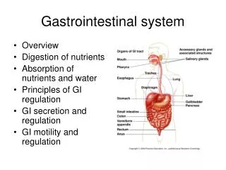
Gastrointestinal system protozoon Giardia lamblia (Giardia intestinalis)
Giardia lamblia ►It is an intestinal flagellate ►Lives in duodenum and jejunum ►Cause “Giardiasis”
Giardia lamblia ► Human pathogen: G. lamblia or G. intestinalis or G. duodenalis, ►Two life stages: trophozoite and cyst. HISTORY ► First observed 1681 by Anthony van Leeuwenhoek
Giardia lamblia ►First observed 1681 by Anthony van Leeuwenhoek ►Described ~200 years later by Vilem Lambl ►First cultured in 1960’s ►Confirmed pathogen 1970’s ►One of most common intestinal parasites ►Causes Giardiasis (beaver fever) ►Geographic region with poor water sanitation
Giardia lamblia ►Species details Single-celled protist 5 species of Giardia ► G. intestinalis/lamblia G. muris in rodents, birds, reptiles G. agilis in amphibians G. ardae in great blue heron G. psittaci in budgerigar
Giardia lamblia ►Morphology www.tulane.edu • Cyst • ► Infective stage in the • environment, • ► Persist in cold water up to • several months • ►Egg-shaped, 8-14µm by 7-10µm • ► Organelle duplication w/out • cytokinesis results • *infour nuclei (Nu) • *four median bodies (MB) • *four axonemes (Ax)
Giardia lamblia ► Morphology www.med-chem.com • Trophozoite • ► Cannot survive in the environment • ► Motile 4 pairs of flagella • ► Pear shaped, bilaterally • symmetrical • ► Relatively flattened, 10-12µm long • ► 5-7µm wide with a large sucking disk on the anterior ventral side • ► Two nuclei