Chapter 21
Chapter 21. Quantitation. Cell Counting Technique. Hemocytometer Electronic Cell Counter. Hemocytometer. Counting chamber (optically flat chamber) Hemocytometer Slide Microscope Cell suspension. Manual Cell Counting. Improved Neubauer. X100. X400. WBC Counting and RBC Counting.

Chapter 21
E N D
Presentation Transcript
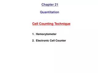
Chapter 21 Quantitation Cell Counting Technique • Hemocytometer • Electronic Cell Counter
Hemocytometer • Counting chamber (optically flat chamber) • Hemocytometer Slide • Microscope • Cell suspension
Manual Cell Counting ImprovedNeubauer X100 X400
WBC Counting andRBC Counting 9大格 25中格 16小格 Cultured Cell Counting = “WBC counting”
http://biology.clc.uc.edu/fankhauser/Labs/Anatomy_&_Physiology/A&P202/Blood/Blood_Counts_practice.htmhttp://biology.clc.uc.edu/fankhauser/Labs/Anatomy_&_Physiology/A&P202/Blood/Blood_Counts_practice.htm
Calculation of cell numbers • Area = 1 mm x 1 mm • Height: 0.1 mm • Average of cell number • Calculation (cell numbers)
Calculation 50 mL 15 mL 1 mL 20 uLcell suspension 200uL Hemocytometer Assume: 28+30+35+56 • Average = 37.25 • Chamber volume =1x1x0.1 = 0.1 • Cell number = (mm3) =1x10⁻⁴ (cm3) 37.25 (cells)÷0.0001 (mL) x15 (mL) TOTAL cell number !!! Various Cell Density !!!
Attention • You have a single-cell suspension • It requires a minimum of 1x10⁶ cell/mL • Do not allow the cell time to settle or adhere in the tip of the pipettebefore transferring them to the chamber • If cell aggregation can not be eliminated, lyse the cells in 0.1M citric acid containing 0.1% crystal violet at 37C for 1 hour and thencount the nuclei
Electronic Cell Counter • Cell sizing • Discrimination between Viableand nonviable cells • Single cells and aggregates Beckman Scharfe System
Electrical Resistance • As each cell passes through theorifice, it changes the resistance tothe current flowing • The size of the pulse is proportionalto the volume of the cell
Automated Cell Viability Analysis (a) Automation of the standard trypan blue assay (b) % Viability (c) Total cell concentration (d) Total viable cell concentration (e) Mean cell size (f) Real time cellular images
Cell Viability 1. Prepare a cell suspension of the cells to be assayed 2. Prepare a 1:1 dilution of the suspension using a 0.4% trypan blue solution. 3. Load the counting chambers of a hemocytometer with the dilution. 4. Let sit for 1-2 minutes (do no leave longer as viable cells may die and begin to take up the dye). 5. Count the number of stained cells and total number of cells 6. The calculated percentage of unstained cells will represent the percentage of viable cells. (within 1 min)
Trypan blue exclusion assay http://www.biomedcentral.com/1471-2202/6/40/figure/F1
MTT ssay MTT (3-(4,5-Dimethylthiazol-2-yl)-2,5-diphenyltetrazolium