Immunostained Kidney Sections of T-Cells and DCs in Obstructed Kidneys
10 likes | 87 Views
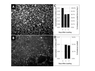
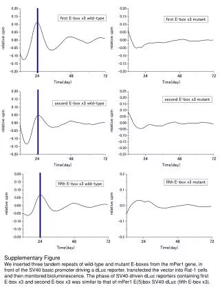
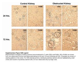
Illustrative examples of kidney sections immunostained for T-cells (CD3) and DCs (F4/80) in non-obstructed and obstructed kidneys at 24 and 72 hours post-left UUL, showcasing staining patterns in the cortex. Positive stained cells are indicated by circles and clusters by ovals.

Immunostained Kidney Sections of T-Cells and DCs in Obstructed Kidneys
E N D
Presentation Transcript
Supplementary Figure S2B Legend Representative examples of kidney sections immunostained for T-cells (CD3) and F4/80+ DCs (F4/80) are shown from non-obstructed (Control) and obstructed kidneys at 24 and 72 hours following left UUL. Examples are shown of staining patterns in the cortex. For CD3-stained sections, individual positively stained cells are highlighted by small circles and clusters of positively-stained cells (72 hour obstructed only) by large ovals.