
DIGESTIVE SYSTEM
DIGESTIVE SYSTEM. Digestive System. muscular tube digestive tract GI tract gastrointestinal tract alimentary canal runs from oral cavity pharynx, esophagus, stomach, small & large intestines, rectum to anus includes accessory organs teeth tongue salivary glands liver gall bladder

DIGESTIVE SYSTEM
E N D
Presentation Transcript
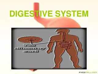
Digestive System • muscular tube • digestive tract • GI tract • gastrointestinal tract • alimentary canal • runs from oral cavity pharynx, esophagus, stomach, small & large intestines, rectum to anus • includes accessory organs • teeth • tongue • salivary glands • liver • gall bladder • pancreas
Functions • provides fuel to keep cells running • provides building blocks for growth & repair • removes residue • protective function for bacteria • largest immune organ
Functions • four integrated steps • Ingestion • intake of food • Digestion • mechanical • physical manipulations • teeth tear & crush • stomach mixes & churns • chemical breakdown • physically manipulated materials broken into smaller fragments by enzymes & acids • Absorption • uptake of nutrients • movement of organic substances, ions, vitamins & water into blood • Defecation • excretion of undigested residue • removal of waste products
Histological Organization • located in peritoneal cavity • lined by serous membrane • visceral peritoneum covers organs • parietal peritoneum lines inner body wall surface • 4 layers • mucosa • submucosa • muscularis externa • serosa
Layers of Wall • Mucosa • innermost epithelial lining • consists of inner epithelium & thin layer of smooth muscle-muscularis mucosae • Epithelium-simple columnar in most of tract • from oral cavity through esophagus & lower anal canal-stratified • Submucosa • loose connective tissue containing blood & lymph vessels with nerve plexus • Muscularis externa • 2 layers of smooth muscle arranged in inner, circular layer & outer, longitudinal layer • important in mechanical processing & movement of materials along tract. • lining of muscularis externa is thrown into foldsincreases surface area • Serosa • inner layer of loose connective tissue • adipose tissue & outer epithelial layer
Nervous System Control of Digestive Functions • sympathetic stimulation inhibits gastrointestinal secretion, motor activity & contraction of gastrointestinal sphincters & blood vessels • parasympathetic stimuli stimulate these
Nervous Control of Digestive Functions • digestive system has its own, local nervous system • enteric nervous system • can function independently of CNS • regulates motility, secretion & blood flow in tract • has more neurons than spinal cord • comprised of two nerve networks • submucosal plexus in submucosa • myenteric plexus between two layers of muscularis externa
Motility in Digestive Tract • Peristalsis • progressive contraction of circular & longitudinal muscles • propels bolus along tract • circular muscles contract behind bolus while circular muscle ahead relaxes
Motility in Digestive Tract • Segmentation • contraction & relaxation of non-adjacent segments of tract • moves contents forwards & backwards • mixes & churns bolus • breaks into fragments • mixes with intestinal secretions
Ingestion • eating • begins food processing • begins chemical & mechanical digestion • oral or buccal cavity • responsible for analysisof material prior toswallowing • mechanical processingvia tongue & teeth • lubrication-mixing ingested material with saliva&mucus • limiteddigestion of carbohydrates & lipids • lined with stratified squamous epithelium • roof-hard & soft palate • floor-tongue
Tongue • mechanicalprocessing • manipulation to assist chewing • sensory analysis • touch, taste, temperature • secretion of linguallipase • beginslipidbreakdown
Salivary Glands • make saliva • controlled by ANS-parasympathetic nervessalivary reflex • Lubricating • Moistening • Parotid • serous, watery secretion containing salivary amylase(starchmaltose) • Submandibular • secretion contains mucus & amylase • Sublinguals • mucus secretion • buffer & lubricant
Teeth • aid in mechanical digestion by mastication or chewing • breaks down connective tissues in plant fibers & meat • helps saturate materials with salivary secretions & enzymes • permits easier deglutition • during mastication, salivary glands secrete salivasoften food into a bolus (semi-solid lump)
Swallowing-Deglutition • involves over 22 muscles in mouth, pharynx & esophagus • controlled by swallowing center in medulla & pons • occurs in three phases • Buccal • Pharyngeal • Esophageal
Buccal Phase • voluntary • tongue pushes just formed bolus toward oropharynx • bolus stimulates tactile receptors • activates next phase
PharyngealPhase • tactile receptors send impulses to deglutition center in medulla • impulses returning from center cause soft palate & uvula to more upward closing off nasopharynx- prevents food from entering nasal cavity • epiglottis covers glottis-opening to larynx • bolus driven downward by constriction of upperthen middle then lower pharyngeal constrictors • as bolus slides into esophagus
Esophageal Phase • esophagus stretchestriggers peristalsis pushes bolus ahead of it • peristalsis carries bolus from upper esophageal sphincter through esophagus to lower esophageal sphincterstomach
Stomach • bolus passes through lower esophageal sphincter into stomach • expanded section of digestive tube between esophagus & small intestine
Stomach Functions • bulk storage • stores ingested food in upper part • digestion • mechanical breakdown of ingested food-lower parts • disrupts chemical bonds by acids & enzymes • produces intrinsic factor • for vitamin B12 absorption
Stomach Parts • Cardia • smallest region • contains mucous glands • protects from stomach acids & enzymes • Fundus • makescontact with diaphragm • Body • largest region • mixing tank • contains gastric glandsacids & enzymes • Pyloris • leads into duodenum of small intestine • inside empty stomach mucosa & submucosa are thrown into folds called rugae • distended when food is in stomach • allow stomach to expand
Stomach Wall • covered with simple columnar glandular epithelium • gastric mucosa is covered with numerous small holes • openings of gastric pits • two or three tubular glands open into bottom of each gastric pit • gastric glands • secrete mucus • secrete acid & enzymes
Gastric Gland Cells • Mucous Neck Cells • Parietal • Chief • Enteroendocrine
Parietal Cells • secrete hydrochloric acid • assists in break down of food • not made in cytoplasm • too strong • would dissolve secretory vessel & destroy cell • H+ & Cl- are made & then secreted out of cell & assembled • maintains pH between 0.8 & 2.0 • kills microorganisms • breaks down plant cell walls & connective tissue in meats • essential for pepsin • converts pepsinogen to pepsin • make intrinsic factor • needed for absorption of vitamin B12
Cells of Gastric Pit • Chief Cells • secrete pepsinogen • inactive precursor of pepsin • digests proteins • Enteroendocrine Cells-G cells • make gastrin
Absorption in Stomach • little absorption • aspirin & ethanol • absorption does not occur because • cells are covered by mucus blanket & therefore never contact chyme directly • cells do not have transport mechanisms needed to absorb materials • gastric lining is impermeableto water • digestion has not been completed • digested food pieces too big
Digestion in Stomach • mechanical digestion-churns bolus & mixes it with digestive juices • chemical digestionbreaks bonds • food digests in stomach for several hours • preliminary digestion of proteins by pepsin • not completed • limited time substances are in stomach • pepsin attacks only specific types of peptide bonds • digestion of carbohydrates & lipids by salivary amylase & lingual lipase • enzymes continue to digest until pH falls below 4.5
Chyme Formation • bolus + secretions soupy mixture-chyme • each peristaltic wave delivers a bit of chyme to small intestine through pyloric sphincter-gastric emptying • chemical digestion in small intestine depends on activity of pancreas, liver & gall bladder-accessory digestive organs
Pancreas • lies posterior to greater curvature of stomach • exocrine acini cells secrete 200-1500 ml of juice/day • secreted into small ducts-unite to form larger ducts- pancreatic & accessory • pancreatic duct joins common bile duct from liver & gall bladder • enters duodenum as hepatopancreatic ampulla • passage of pancreatic juice & bile through this into small intestine is controlled by sphincter of the hepatopancreatic ampulla or Sphincter of Oddi.
Pancreatic Juice • mixture of water, salt, enzymes, zymogens & sodium bicarbonate • secretions controlled by hormones of duodenum • chymeduodenum secretin pancreas watery buffer pH 7.5-8.8 raises pH of chyme • chymeduodenumCCK pancreas pancreatic enzymes
Pancreatic Secretions • pancratic amylase • starch breakdown • ribonuclease & deoxyribonuclease • nucleic acid breakdown • pancreatic lipase • lipid breakdown • zymogens: trypsinogen & carboxypeptidase • a brush border enzyme-enterokinase cleaves trypsinogentrypsin • trypsin then works on other inactive precursorsactive ones
Liver & Gall Baldder • accessory digestive organs • liver-inferior to diaphragm • gall bladder-in a depression on posterior surface of liver • Liver-two principle lobes-a larger right & a smaller left lobe • connected by a mesentery fold-falciform ligament • right lobe includes an inferior quadrate & a posterior caudate lobe
Liver & Gall Bladder • hepatocytes-major functioning cells of liver make 800 -1000 mls of bile each day • bile leaves liver via right & left hepatic ducts which unite as common hepatic duct • these join with the cystic duct from the gall bladder to form the common bile duct • bile is stored &modified in the gall bladder • enters small intestine via cystic duct • does not enter small intestine until gallbladder contracts • principal stimulus for release- cholecystokininCCK
Emulsification • bile contains water, bile salts, bile pigments, cholesterol, lecithin & several ions • bile salts are important in digestion of lipids • lipids are not water soluble • mechanical processing results in large drops • bile salts breaks down large lipid globules into a suspension of smaller lipid globules • process called emulsification • increases surface area available for enzymatic attack • digested lipids are absorbed in lacteals of the small intestine
Liver Functions • carbohydrate metabolism • stabilizes blood glucose by glycogenolysis & gluconeogenesis • lipid metabolism • removes lipids for storage or breaks down lipids when needed • amino acid metabolism • removes excess amino acids • removes waste products • amino acidsammonia • neutralizes ammonia by converting it to urea • important in drug inactivation • vitamin storage • fat soluble vitamins-A, D, E & K & B12 • mineral storage • stores iron bound to ferritin • phagocytosis & antigen presentation • Kupffer cells engulf old RBCs, debris, etc, & stimulates immune system • synthesis of plasma proteins
Small Intestine • about 20 feet long • 90% of nutrient absorption • Duodenum • next to stomach • mixing bowl • Jejunum • bulk of chemical digestion & absorption • Ileum • longest part • ends at ileocecal valve • sphincter controlling release of substances into large intestine
Small Intestine Lining • folded into transverse folds-plicae • permanent • increase surface area for absorption • covered by simple columnar epithelium • microvilli project from cells of epithelium forming brush border • increase surface area more • allow chyme to contact more of small intestine wall • increased contact means more efficient food absorption Brush Border
Small Intestine Epithelium • absorptive cells • digest & absorb nutrients in chyme • goblet cells • make mucus
Intestinal Villi • mucosa is thrown into folds forming intestinal villi • Core contains lymph vessel- lacteal • absorbs products of fat digestion • at base-entrance to intestinal glands- crypts of Lieberkuhn • secrete1-2 liters of intestinal juice each day • produce brush border enzymes • Paneth cells • secrete lysozyme • Enteroendocrine cells • S cells • make secretin • CCK cells • make cholecystokine • K cells • make GIP-glucose-dependent-insulinotropic peptide
Duodenum • contains duodenal or Brunners glandsmucous • primary function of duodenum • receive chyme & neutralize acids
Ileum • contains Peyer’spatches • aggregates of lymphoid nodules • protection from bacteria
Movement in Small Intestine • as chyme enters duodenumperistaltic contractions (migrating motility complexes) move it toward jejunum • segmentation-contraction & relaxation of non-adjacent segments of the tract mixes & churns the material breaking it into fragments & mixing it with intestinal secretions
Chemical Digestion & Absorption • begins in mouth • salivary glandssalivary amylase • polysaccharides broken into di- & tri- saccharides • salivary amylase & pancreatic amylase continue to break down in stomach • a brush border enzyme-α dextrinase clips off one glucose at a time making di & tri saccharides • monosaccharides are made by brush border enzymes specific for specific disacchardies • maltase splits maltose • sucrase splits sucrose • absorbed by facilitated transport
Chemical Digestion &Absorption • fat digestion begins in mouth-lingual lipase • continues in stomach • fat enters small intestine as coarse emulsion created by lipase digestion & mechanical mixing • lipid droplets too big to be absorbed or further broken down • bile salts (phospholipids) coat emulsion stabilizing fat droplets • pancreatic lipase digests triglycerides in emulsion • triglyceridesmonoglycerides & free fatty acids • absorbed by simple diffusion into lacteals
Chemical Digestion & Absorption • protein digestion begins in mouth • mechanical processing • chemical processing • takes place in stomach with HCl • pepsin continues digestion in stomach breaking peptide bonds • other proteases & peptidases found on brush border of intestinal villa cells-trypsin, elastin, chymotrypsin continue to breakdown protein bonds • carboxypeptidases break off individual amino acids from ends of peptides • absorbed via facilitated diffusion
Large Intestine • begins at end of ileum • ends at anus • attached to posterior abdominal wall by mesocolon • ileocecal sphincter regulates passage of chyme from the small to the large intestine • relaxes when food leaves stomach-gastroileal reflex • cecum-first part • hangs inferior to ileocecal valve • attached-appendix • functions: reabsorbs water • compacts intestinal contents into feces • absorbs vitamins • stores fecal materials
Large Intestine-Colon • colon-largest part • ascending colon • transversecolon • descending colon • sigmoid colon • muscularis layer has inner circular layer & longitudinal muscle layerconcentrated into 3 bands called teniaecoli • contraction of these pull wall into bulging pockets or haustra • permits expansion & elongation of colon


