Download

1 / 22
240 likes | 350 Views
Learn about the pathophysiology, risk factors, common uropathogens, diagnostic tests, and specimen collection methods for urinary tract infections. Understand the importance of routine urine culture and how to interpret the results accurately.

E N D
Pathophysiology of urinary tract infection • Ascending route of infection is the most common. • Hospital infection associated with lower urinary tract instrumentation (catheterization, cystoscopy). • Once in the bladder uropathogens multiply, then pass up the ureters to the renal pelvis and parenchyma. • Source of uropathogens: enteric bacteria mainly.
Urinary tract infection more common in women than men • Short female urethra. • Close proximity to perianal areas. • Men also have an antibacterial substance in their prostate gland that reduces their risk.
Pathophysiology of urinary tract infection • Cystitis: (lower urinary tract infection) • Pyelonephritis: infection of the kidney with acute suppurative inflammation of the • pelvis, • medullary and cortical tubules, • corticomedullary intersititum • Urosepsis: bacteremia due to pyelonephritis • Papillary necrosis • Sloughing of necrotic pyramids • Perinephric abscess
Risk factors in complicated urinary tract infection • Indwelling catheters • Urinary calculi • Neurogenic bladder • Prostatic enlargement • Uterine prolapse • Urologic instrumentation or surgery • Renal transplantation • Diabetes mellitus
Common uropathogens • Escherichia coli • Other Enterobacteriaceae (Klebsiella, Enterobacter, Proteus, Citrobacter) • Pseudomonas aeruginosa • Enterococcus • Staphylococcus saprophyticus • Staphylococcus aureus • Associated with staphylococcemia • Streptococcus agalactiae (group B) • Denotes vaginal colonization in pregnant women • Candida
Uncommon uropathogens • Corynebacterium urealyticum • Haemophilus influenzae and H. parainfluenzae • Blastomyces dermatitidis • Neisseria gonorrhaeae • Mycobacterium tuberculosis
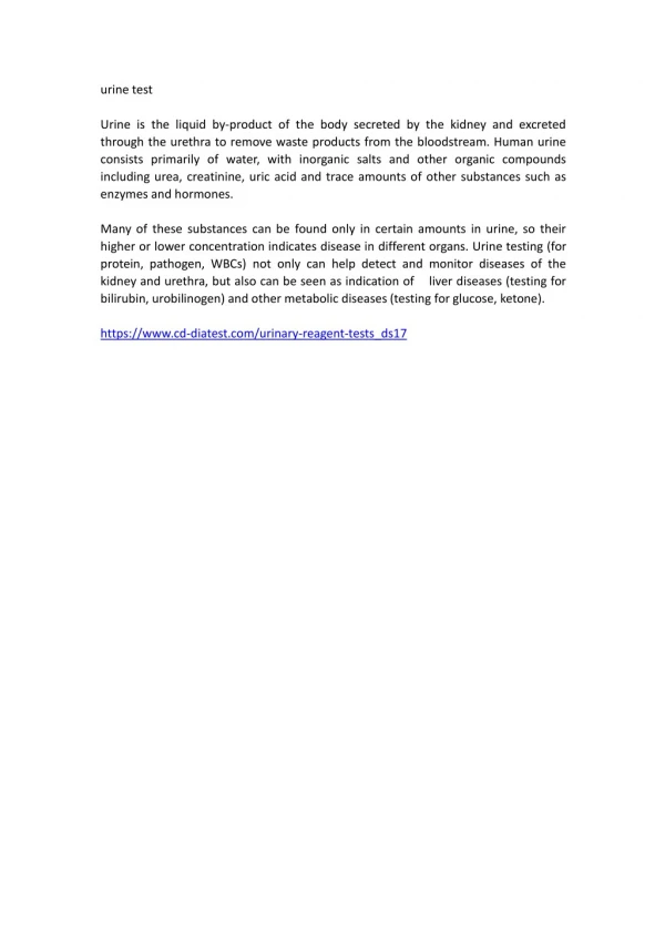
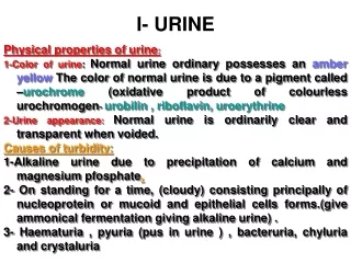
Notes • Adult urine volume = 600 – 2500 ml /24hr. • Oligouria: marked decrease in urine flow < 400 ml/24hr. • Polyuria: Marked increase in urine flow > 2500 ml/24hr. • Anuria: less than 100 ml/24hr. • Nocturia: excessive urination during night.
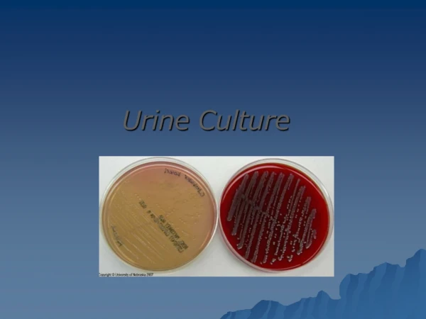
Routine Urine Culture • Aim of the test • An etiological diagnosis of bacterial urinary tract infection by quantitative • Cultivation of the urine with identification and susceptibility test of the isolated bacteria(s). • Criteria of specimen rejection • Un-refrigerated specimen or specimen older than 2 hours may be subject to overgrowth. • Unlabeled specimen; mislabeled specimen. • Specimen in expired transport container. • 24 hours urine specimens.
Types of urine specimens • First-voided morning urine optimal (generally bacteria have been proliferating in bladder urine for several hours) • Midstream urine specimens (initially voided urine contains urethral commensals) • Indwelling catheters (freshly placed, urine aspirated by needle inserted into catheter) (Foley catheter tips not acceptable) • Straight catheter specimens • Suprapubic aspirates (infants or children, recovery of anaerobes) • Cystoscopic collection of urine
Pre specimen processing • Urine collected in sterile specimen container must be processed within hours, or refrigerated and processed within 24 hours. Who will collect the specimen • Midstream urine is collected by the patient. • If disabled, nursing staff will assist in collection. • For catheterized specimen, nursing staff will collect the specimen. • Suprapubic aspiration is performed by the physician.
Catheterized urine • Cleanse periurethral area with soap and water • Insert catheter into bladder • Discard initial urine • Collect specimen in sterile cup • Chronic indwelling Foley catheter • Clamp tubing below junction (or port) • Disinfect with alcohol • Insert needle (on syringe) through port or catheter wall and aspirate. • DO NOT recontamination
Quantity of specimen To fill line in transport tube (~20 mL). Time relapse before processing the sample The maximum time allowed for processing a urine sample is 2 hours from the time of collection. Storage At room temperature unless delay is inevitable; it must be refrigerate or mixed with preservative like boric acid.
Inoculation of urine • Inoculation of urine for quantitative culture (colony forming units→ CFU’s) performed with a calibrated 0.001 mL(1µL) and 0.01 mL (10µL) plastic or wire loop • Nutrient agar or sheep blood agar utilized for quantitative urine culture • With 0.001 ml loop, 1 colony on SBA equivalent to 1,000 CFU’s per mL of urine • With 0.01 ml loop, 1 colony on SBA equivalent to 100 CFU’s per mL of urine • MacConkey agar utilized as selective differential agar for gram-negative bacteria
Initial report The use of dipstick designed to detect the presence of urine nitrite and to indirectly estimate the number of segmented neutrophiles through the detection of leukocyte esterase activity. Rationale for the nitrate test is that most urinary tract infections are caused by nitrate reducing members of the family Enterobacteriaceae. Pyuria: the increased number of WBC in urine sample.
Interpretation: Clean Voided Specimen • Normal: <10,000 organisms per ml. • A plate count of 100,000 CFU/ml of pure culture should be considered positive, and isolated organism should be identified and sensitivity test will be performed. • A plate count between 10,000 – 100,000 CFU/ml is considered suspected . • A plate count less than 10,000 CFU/ml is considered negative.
Post specimen processing • Interfering factors: • Patient on antibiotic therapy. • Improper sample collection. • Result reporting: • Report wet mount as an initial report. • Report the isolated pathogen and its sensitivity pattern as a final report. • Turn around time: • Wet mount results should be available 1 hour after specimen receipt. • Isolation of a possible pathogen can be expected after 2-3 days. • Negative culture will be reported out 1-2 days after the receipt of the specimen.