Download

1 / 83
880 likes | 1.15k Views
Urine Analysis. Importance of urine analysis. It can detect diseases which pass unnoticed. For example D.M, chronic UTI. Diagnosis of many renal diseases. As nephrotic, nephritic syndrome, acute renal failure, multiple myeloma. Nephrotic syndrome vs Nephritic syndrome. Nephrotic syndrome:

E N D
Importance of urine analysis • It can detect diseases which pass unnoticed. For example D.M, chronic UTI. • Diagnosis of many renal diseases. As nephrotic, nephritic syndrome, acute renal failure, multiple myeloma
Nephrotic syndrome vs Nephritic syndrome Nephrotic syndrome: • 1. Massive proteinuria2. Hypoalbuminemia3. Edema4. Hyperlipidemia/hyperlipiduria Nephritic syndrome: • 1. Hematuria2. Oliguria3. Azotemia4. Hypertension
Urine Composition Urine, a very complex fluid, is composed of 95% water and 5% solids . It is the end product of the metabolism carried out by billions of cells and results in an average urinary out put of 1-1.5 L per day. Urine may also contain formed elements such as cells, casts, crystals, mucus and bacteria. Almost all substances found in urine are also find in the blood although in different concentration.
Renal Physiology Three basic renal processes Glomerular filtration. Tubular reabsorption. Tubular secretion.
Glomerular filtration • Glomerulus filters incoming blood, all substances except cells and large molecules pass into further sections of the nephron. • Filtration process requires adequate pressure. • Water, electrolytes, glucose, amino acids, urea, creatinine pass freely and enter the proximal tubule. • If 200 liters of filtrate enter the nephrons each day, but only 1-2 liters of urine result, then obviously most of the filtrate (99+ %) is reabsorbed.
Reabsorption • can be active or passive, and occurs in virtually all segments of the nephron. • Renal threshold for each substance determines whether it is reabsorbed or secreted. However, some substances have no renal threshold e.g H2O. • Glucose, actively reabsorbed in the proximal tubules according to the renal threshold • Na, actively reabsorbed according to the diet.
Secretion • It is the reverse of reabsorbtion. • It is either by active process or by diffusion. • H +,K+, ammonia. Are the principle particles that is exsecreted by the kidney. • H+ ions play an important role in acid base balance.
Control Of Urine Excretion • Antidiuretic Hormone (ADH) • Aldosterone
Water Balance Water loss is under the control of ADH. ADH responds primarily to changes in osmolality and intravascular volume. Increased osmolality stimulates ADH secretion which increases the permeability of collecting tubules to water resulting in more concentrated urine. In dehydration, reabsorption of water is increased, In states of water excess, tubules reabsorb water at only a minimal rate resulting in excretion of large volume of dilute urine.
Specimen Collection The specimen must be collected in a clean dry, disposable container. The container must be properly labeled with the patient name, date, and time of collection. The labels should be applied to the container and not to the lid. The specimen must be delivered to the laboratory on time and tested within 1hr, OR it should be refrigerated or have an appropriate chemical preservative added. eg. (Toluene, formalin or boric acid).
Changes in unpreserved urine Transformation of urea to ammonia which increase pH. urease Urea 2NH3 + Co2. (Bacteria) Decrease glucose due to glycolysis and bacterial utilization. Decrease ketones because of volatilization. Decrease bilirubin from exposure to light. Decrease urobilinogen oxidation to urobilin. Increase bacterial number. Increase turbidity caused by bacteria & amorphous. Disintegration of RBCs casts, particularly in diluted alkaline urine. Increase nitrite due to bacterial reduction of nitrate. Changes in color due to oxidation or reduction of metabolic.
Types of specimens Random specimen (at any time). First morning specimen 24 hr’s collection Post. Prandial sample Clear catch sample (midstream urine) Catheterized urine Supra - pubic
urinanalysis • Macroscopic • Chemical • microscopic
Physical examination of Urine (Macroscopic, Gross)
Physical Examination of Urine Visual examination of physical characteristics • Color • Turbidity • Odor • Volume • Specific gravity
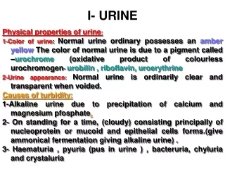
Appearance (color and clarity) Color: Normal urine color has a wide range of variation ranging from pale yellow, straw, light yellow, yellow, dark yellow amber due to urobillin,trace of urobilinogen appears in urine The color is affected by: Concentration of urine pH Metabolic activity. Diet intake (Beet). Drugs may change urine color.
Abnormalities in color Colorless or pale yellow: High fluid intake Using of diuretic. Diabetes Mellitus. Diabetes Insipidus. Alcohol ingestion Dark yellow: Low fluid intake. Excessive sweating Dehydration (burns, fever). Carrots or vitamin (A) orange urine. Antibiotic used against E. coli in urinary tract infection).
Brownish yellow: • Bilirubin on shaking yellow foam will appear. • Urobilin on shaking the foam has no color. • Yellow – green • Bilirubin Biliverdin (greenish). • Which give a yellow foam & (- ve) test for bilirubin. • Blue – Green: • Pseudomonas Infection. • Black Urine: • Alkaptonurea, a disease of tyrosine metabolism.
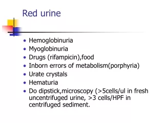
Pink – Red: Due to the presence of fresh blood or Hb, fresh blood will give smoky color while Hb gives clear reddish urine, which may be due to: - Urinary tract infection Calculi Trauma Menstrual contamination. Dark brown: Methemoglobin if bloody sample long standed, Hb will be oxidized. Malignant Melanoma light Melanogen (Colorless) Melanin (Brown).
Clarity (Transparency). Normal urine clear or transparent, any turbidity will indicate. WBCs (pus). RBCs Epithelial cells Bacteria Casts Crystals Lymph Semen.
odor Fresh normal urine has a faint aromatic odor due to the presence of some volatile acids. In some pathological conditions, certain metabolites may be produced to give a specific odor such as: Fruity odor is due to acetone Diabetic urine Ammoniac odor urine standing long time Offensive odor Bacterial action of pus (UTI). Apple odorAsparagus Mousy odor Phenylalanine (phenyl keto urea “PKU” ).
d. Volume Adult urine volume = 600 – 2500 ml /24hr. Children urine volume = 200 – 400 ml /24hr. (4ml / kg / hr). Which depends on: Water intake. External temperature. Mental and physical state. Intake of fluid and diuretics (Drugs, alcohol – tea).
Abnormalities Oligouria: marked decrease in urine flow < 400 ml/24hr. Polyuria: Marked increase in urine flow > 2500 ml/24hr. Anuria: less than 100 ml/24hr. Nocturia: excessive urination during night. Causes of polyuria: Increased fluid in take. Increased salt intake and protein diet, which need more water to excrete. Diuretics intake (certain drugs, drinks). Intravenous saline or glucose. Diabetes Mellitus. Diabetes Insipidus. Renal disease. Hypoaldasteronism.
Causes of Oliguria: Prerenal: In response to hypoperfusion of the kidney (e.g. as a result of dehydration by poor oral intake, cardiogenic shock, diarrhea, massive bleeding) Renal: Due to kidney damage (Calculi, tumor, severe hypoperfusion, medication) Post renal: As a consequence of obstruction of the urine flow (e.g. enlarged prostate, tumour compression urinary outflow, expanding hematoma or fluid collection) Causes of anuria: Sever Renal Defect and loss of urine formation mechanism. Due to the presence of stone or tumor. Post transfusion hemolytic reaction.
e. Specific Gravity Specific gravity (which is directly proportional to urine osmolality which measures solute concentration) measures urine density, or the ability of the kidney to concentrate or dilute the urine over that of plasma. Specific gravity between 1.002 and 1.035 on a random sample should be considered normal if kidney function is normal.
Low specific gravity • Diabetes Insipidus. • Excessive water intake. • Glamerulonephritis. • Sever renal damage. • High specific gravity: • Diabetes mellitus. • Nephrosis. • Fever since urine is conc. • X ray contrast media.
Measurement of spg Urinometer: which is consists of a weighted float a hatched to a scale that has been calibrated in terms of urine spg. (1.00 – 1.040) the weighted float displaces a volume of liquid equal to its weight and has been designed to sink to a level of 1.000 in distilled water. Disadvantages of urinometer: The minimum amount of urine to be measured is about 15 ml. If the urine is so turbid it is difficult to read the result.
Refractometer Determine spg by measuring the refractive index of urine Reagent strip: Which contain polyelectrolyte, when ions increase in urine, more acidic groups are released, the change in pH will take place which change the color of bromothymol blue indicator.
Acid-Base Equilibria The kidneys role in controlling body pH is accomplished by preserving HCO3– and removing metabolic acids. Regeneration of HCO3– HCO3 –are filtered by the glomerulus. HCO3–combines with H+ in the lumen of renal tubules to form H2CO3. H2CO3 is degraded to CO2 + H2O.
CO2 diffuses into proximal tubules and is converted to H2CO3 by the action of carbonic anhydrase, then it is degraded back to H+ and HCO3. This regenerated HCO3 is transported into the blood to replace the depleted one by metabolism, H+ are secreted into the tubular lumen and enter the urine. NH3 NH3 is formed in the renal tubules as a result of glutamine deamination by glutaminase, NH3 then react with H+ to form NH4 which is excreted in urine.
PH One of the important functions of the kidneys is pH regulation, the glomerular filtrate of blood plasma is usually acidified by renal tubules and collecting ducts from a pH of 7.4 to about 6 in the final urine to keep blood pH about 7.4. Hence, urine pH must vary to compensate for diet and products of metabolism, this function takes place in the distal convoluted tubule with the secretion of both H+ & NH3+ and reabsorption of bicarbonate. Normal urine pH is (4.6 – 8.0) as average (6.0)
Clinical significance of pH Determine the existence of metabolic acid base disorder. Precipitation of crystals to from stone requires specific pH for each type Hence, pH control may inhibit the formation of these stones by control diet. Crystals found in alkaline urine: Ca carbonate, Ca phosphate, Mg phosphate, and amorphous phosphate. Crystals found in acidic urine: Ca oxalate, Uric acid, Cystine, Xanthine and amorphous urate. May indicate the presence of urinary tract infection caused by urea splitting organisms. urease Urea NH3 + CO2.
Defects in renal tubular secretions and reabsorption of acid & base. Determination of unsatisfactory specimens.
Test for pH Reagent strip which has an indicator (methyl red – bromothymol blue indicator) or other indicators. Alkaline urine is found in: Patient with alkalemia, UTI, diets high with citrus fruits or vegetables. Acidic urine is found in: Patient with acidemia, starvation, dehydration, high diets with meat products
Chemical properties of urine
A small amount of protein (50 – 150 mg / 24 hrs) appears daily in the normal urine. More than 150 mg/day is defined as proteinuria. This amount of protein is form of: 40% consist of albumin, which may escape from the glomerulus membrane & not reabsorbed. 40% of mucco-protein which is secreted from the renal tubule and other secretions from genitalia. 20% other traces of non-plasma proteins. Proteinuria: The presence of detectable amount of proteins in urine. Protein
Causes of proteinuria Glomerular membrane damage,which may be: Primary: due to primary glomerular defect as glomerulnephritis Secondary: - due to external disease that affects the glomerular function as: 1- SLE 2- Drug 3- Septicemia Prerenal Proteinuria : - Over flow / over load, increase of LMW protein such as multiple myeloma ex. Bence Jones protein. Tubular proteinuria: Presence of LMW protein, absorption problems. Functional or Nonpathogenic proteinuria due to: Fever, Emotional, Cold, Later months of pregnancy, Postural (as long standing & exercises).
Tests for protein Dipstick: when an indicator dye is adsorbed to protein the paper spot in the dipstick is impregnated with citrate buffer (PH = 3.0) containing Bromphenol blue, which is most sensitive to albumin but detects globulins and Bence-Jones protein poorly. Not: Bromphenol blue is yellow at pH 3.0 and blue at pH 4.2, at pH (3).
The color is compared with that of the protein content from (30 –1000mg /dl).
Precipitation tests Heat denaturation for protein precipitation. Sulfosalicylic acid (more sensitive) Test for bence – Jones protein First heat the urine between 40 – 60 ْC, precipitation will occur then continue heating till 100 ْ C so the precipitation will disappear (clear). If you cool the urine till 40 – 60 ْC the precipitation will occur again.
Combined use of dipstick and sulfosalicylic acid • If both are +ve then proteinuria is present. • If dipstick 1+ and sulfosalicylic negative then there is probably no pathologic concentration of protein. • If dipstick negative and sulfosalicylic positive then the protein may be Bence Jones protein should confirmed by immunologic method.
Glucose Under normal conditions, all most all of glucose filtered by glomerulus is reabsorbed in the proximal convoluted tubule, by an active process to maintain the plasma concentration of glucose. Less than 0.1% of glucose normally filtered by the glomerulus appears in urine (< 130 mg/24 hr). Threshold substances: Substances that are completely absorbed by the tubules when their plasma concentration is normal and not completely absorbed by the tubules if their plasma concentration exceeds normal levels. The threshold of glucose is 180 mg / dl.
Glycosuria may be due to: Reabsorption defect Increase Blood glucose, in the following cases: Diabetes mellitus Alimentary glycosuria (transitory), after meal. Stress in which elevation of epinephrine leads to increase glycogenolysis, and cortisol increase gluconeogenesis. Pancreatic disease affect insulin-secreting gland. Decrease reabsorption ability.