Urine Luck!
Urine Luck!. Renal slides by Dan Cushman Donations accepted and strongly encouraged. Interlobar artery . Lobe. Cortex. Renal artery. divides into anterior and posterior branches. Medulla. Renal vein. Ureter. Renal pelvis. Kidney parasite Parasitium nephrotium.

Urine Luck!
E N D
Presentation Transcript
Urine Luck! Renal slides by Dan Cushman Donations accepted and strongly encouraged
Interlobar artery Lobe Cortex Renal artery divides into anterior and posterior branches. Medulla Renal vein Ureter Renal pelvis
Kidney parasiteParasitiumnephrotium (Just kidding, of course, it’s a nephron)
Vessels Name the arteries of the kidney from largest to smallest 1. Renal Artery 2. Interlobar artery 3. Arcuate artery 4. Interlobular artery 5. Afferent arteriole 6. Efferent arteriole
Renal corpuscles Kidney vessels Cortex Interlobular vessels Arcuate vessels Medulla Interlobar vessels X10
Nephron segments Proximal convoluted tubule Proximal straight tubule Cortex Descending limb of Henle Thin Ascending limb of Henle Thick Ascending limb of Henle Medulla Distal convoluted tubule Collecting duct
Renal corpuscle (Layer) Visceral layer Efferent arteriole Urinary pole Afferent arteriole (Layer) Parietal layer
What is the best word in nephrology? Corpuscle What is the best structure in nephrology? There really is not an answer to this question. It’s more of a personal reflection question with no objective answer. “Best” is hard to quantify.
Endothelial cell nucleus Filtration apparatus Podocyte Primary process Glomerular basement membrane Capillary lumen Secondary process (pedicel) (Glomerulus lumen) Where are large anions repelled?
What is this? Can’t you read?
Mesangial cells Visceral layer – Podocyte What are the functions of mesangial cells? • Phagocytic – clean the basement membrane, ex. Remove immune complexes from the membrane. • Support – podocytes. • They are contractile- can regulate glomerular lumen. • Secretory – Interleukin-1 and platelet-derived growth factor. These respond to glomerular injury. Efferent arteriole Afferent arteriole Parietal layer
So… tell me about the juxtaglomerular cells. • Smooth muscle cells of the afferent arteriole. They are innervated by sympathetic neurons and secrete renin into the blood. Macula densa Distal convoluted tubule Afferent arteriole Juxtaglomerular cells
Which is which? Proximal Tubule Distal Tubule (Brush Border)
Which is which? Full Bladder Empty Bladder
Which portion turns into a kidney? What is its main signaler? Neural tube Intermediate Mesoderm Paraxial Mesoderm Gut
Order these three chronologically Which one turns into your kidney? Mesonephric kidney Pronephric kidney Metanephric kidney Which one turns into the ductus deferens?
What transcription factor do I create? Mesonephros tubules Metanephros Hindgut Mesonephric duct WT1 Cloaca Metanephric Mesoderm (blastema) Ureteric bud Which induces what other factor? GDNF
Name the defect Horseshoe kidney Pelvic kidney Bifid ureter
Where’s the bladder? Here Where’s the love? All around us
Match each to a line Filtered Excreted Secreted
Will the hypertonic gradient in the medullary interstitium ↑ or ↓?
Match transporters with location Thick ascending loop of Henle Na/HCO3 antiport Thin ascending loop of Henle Na/glucose symport Descending loop of Henle Na/H antiport Proximal Tubule Proximal Tubule Proximal Tubule Na/K/2 Cl symport
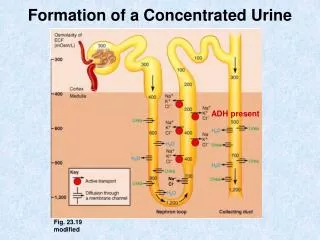
How do V2 receptors function? • They are localized in the basolateral membranes of principal cells. Activation of V2 receptors elevates cyclic AMP in these cells, which leads to insertion of water-permeable channels (AQP2) into the lumenal membrane. What causes ADH release (6)? • Increase in plasma osmolarity (1-2% threshold) • Reduction in circulating blood volume (>10%) and blood pressure • Angiotensin II • Stress (physical or emotional), pain • Nausea • Standing upright (→orthostatic antidiuresis)
What percentage is reabsorbed in each section? Coupled with which anions? 65% Na 65% H2O Cl- (50% of its filtered amount) and HCO3- (90%) 25% Na 15% H2O Coupled with which anions? Cl- (33% of its filtered amount)
Where is the lower O2 content? The cortex The medulla
Order these in terms of reaction speed Baroreceptor reflex Fast Renal control of body NaCl Medium Angiotensin II Slow
Renal Regulation: High, Intermediate, or Low? Intermediate HCO3- • Low [Cr]P ↑ as nephrons are lost Creatinine Urea Low • High Na+ High Water Intermediate Ca2+
Match the lines Which will have the greatest osmolarity? Saline Saline Glucose or water Alcohol
↑/↓ ↑/↓ ↑/↓ ↑/↓ ↑/↓ ↑/↓ ↑/↓
Each caused by what? ↑ perfusion pressure, NE, (ATII) ↑/↓ ↑/↓ ↑/↓ ATII ↑/↓ ↑/↓ ↑/↓ ATII ↑/↓ ↑/↓ ↑/↓
What will increase FENa? Constriction or Dilation Dilation of efferent arteriole Increase or Decrease Decrease activation of RAA system Increase secretion of natriuretic hormones Increase or Decrease
↑/↓ ↑/↓ ↑/↓ ↑/↓ ↑/↓ ↑/↓ ↑/↓ ↑/↓ ↑/↓
Aldosterone Direct stimulants (2): ↑ [K+]plasma, ATII Most importantly, what happens to FENa? Acts on (2): Late distal tubule, collecting ducts It decreases! Principal cells do what (4): • - Increased Na+ permeability of lumenal membrane • - Increased K+ permeability of lumenal membrane • - Increased lumenal Na+/H+ exchange • - Increase in activity and number of basolateralNa+,K+-ATPase pumps Intercalated cells do what (1): • - Increased lumenal H+-ATPase activity
↑/↓ ↑/↓ ↑/↓ ↑/↓ ↑/↓ ↑/↓
↑/↓ ↑/↓ ↑/↓ ↑/↓ ↑/↓ ↑/↓ ↑/↓
Choose: Osmoregulation or Volume Regulation? Osmoregulation Senses plasma osmolality • Osmoregulation Regulates ADH, thirst Affects urine Na excretion Volume Regulation • Volume Regulation Takes days to occur Volume Regulation Edema is a physical sign Volume Regulation ADH, ANP, RAA system
Volume Regulation (Sensor) Baroreceptors ↑ Sympathetics (hormone) ↑ Renin ↑ ATII
Name the three substances that increase the Na/K ATPase activity 1 1 2 2 3 2
What exchange occurs in principal cells? • Na+in, K+ and H+out What exchange occurs in intercalated cells? K+ in, H+ out
↑/↓ ↑/↓ ↑/↓ ↑/↓ ↑/↓ ↑/↓ ↑/↓ ↑/↓
Name the drugs B (3 drugs) A