Download

1 / 33
400 likes | 771 Views
Urine analysis. Macroscopic urinalysis. Is the direct visual observation of the urine, noting its volume, color, clarity or cloudiness, etc Normal urine is typically pale yellow and clear . Obvious abnormalities in the color, clarity, and cloudiness may suggest different diseases.

E N D
Macroscopic urinalysis Is the direct visual observation of the urine, noting its volume, color, clarity or cloudiness, etc Normal urine is typically pale yellow and clear . Obvious abnormalities in the color, clarity, and cloudiness may suggest different diseases.
Normal Urine Abnormal Urine
Dipstick chemical analysis • Urine dipstick is a narrow plastic strip which has several squares of different colors attached to it. • Each small square represents a component of the test used to interpret urinalysis. • Colors generated by each pad are visually compared against a range of colors on brand-specific color charts • The entire strip is dipped in the urine sample and color changes in each square are noted.
The squares on the dipstick represent the following components in the urine Nitrite (suggestive of bacteria in urine) Bilirubin ( possible liver disease or red blood cell break down) Urobilinogen ( possible liver disease)
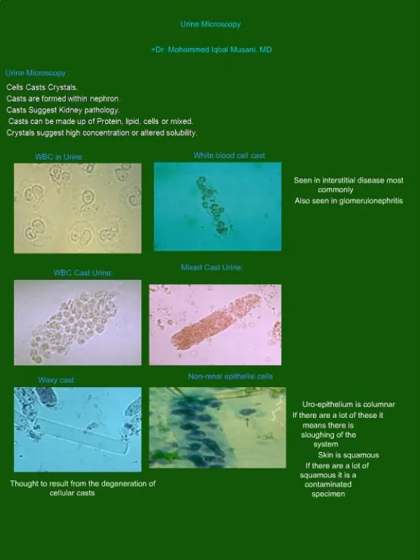
MICROSCOPIC URINALYSIS Microscopic examination used to view elements that are not visible without microscope. e.g cells 1. Red Blood Cells: Hematuria is the presence of abnormal numbers of red cells in urine due to: a. Glomerular damage b. Tumors c. Urinary tract stones d. Upper and lower urinary tract infections
Hematuria Two Types of Hematuria • Gross hematuriameans that the blood can be seen by the naked eye. The urine may look pinkish, brownish, or bright red. • Microscopic hematuria means that the urine is clear, but blood cells can be seen under a microscope.
2.White Blood Cells Pyuria refers to the presence of abnormal numbers of leukocytes that may appear with infection in either the upper or lower urinary tract or with acute glomerulonephritis. Usually, the WBC's are granulocytes WBCs - ≤2-5 WBCs/hpf
3. Epithelial Cells • Renal tubular epithelial cells, contain a large round or oval nucleus and normally slough into the urine in small numbers. However, with nephrotic syndrome and in conditions leading to tubular degeneration, the number sloughed is increased. • ≤15-20 squamous epithelial cells/hpf
4. Casts • Urinary casts are cylindrical structures produced by the kidney and present in the urine in certain disease states. • They are formed in the distal convoluted tubule (DCT) and collecting ducts of nephrons, then dislodge and pass into the urine, where they can detected by microscopy. -Urinary casts may be made up of cells (such as white blood cells, red blood cells, kidney cells) or substances such as protein.
The factors which favor protein cast formation 1. low flow rate of the filtrate 2. high salt concentration 3. low pH all of which favor protein denaturation and precipitation, particularly that of the Tamm-Horsfall protein. Protein casts with long, thin tails formed at the junction of Henle's loop and the distal convoluted tubule are called cylindroids.Hyaline casts (Tamm-Horsfallproteins) can be seen even in healthy people.
Hyaline casts are composed primarily of a mucoprotein (Tamm-Horsfall proteins) secreted by tubule cells. The Tamm-Horsfall protein secretion (green dots) is illustrated in the diagram below, forming a hyaline cast in the collecting duct
Red blood cells may stick together and form red blood cell casts. Such casts are indicative of glomerulonephritis, with leakage of RBC's from glomeruli White blood cell casts may also be present with glomerulonephritis. Their presence indicates inflammation of the kidney, because such casts will not form except in the kidney.
RBCs Crenated RBC
Bence Jones proteins Bence Jones proteins are small proteins found in the urine. Testing for these proteins is done to diagnose and monitor multiple myeloma and other similar diseases. Bence Jones proteins are considered the first tumor marker. A tumor marker is a substance, made by the body, that is linked to a certain cancer, or malignancy. Bence Jones proteins are made by plasma cells, a type of white blood cell. The presence of these proteins in a person's urine is associated with a malignancy of plasma cells.
Bence Jones protein cast (myeloma cast) from the urinary sediment of a patient with lambda-Bence Jones type multiple myeloma. Sternheimer stein, X200
Pregnancy tests • It detects a hormone in the body called human choronicgonadotropin (hCG). • hCGis a hormone produced during pregnancy. It appears in the blood and urine of pregnant women as early as 10 days after conception • hCGis released into the body by the placenta when a woman is pregnant.
The urine hCG test is usually performed by placing drops of urine on a prepared chemical strip. It takes 1-2 min. for a result.