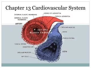
Cardiovascular System Chapter 13
Cardiovascular System Chapter 13. Objectives: Identify structures and functions of the cardiovascular system. Trace the flow of blood through the body. Coverings of the Heart. Pericardium : (?) Visceral (?) pericardium (“epicardium” (?) ) Innermost layer Covers the heart

Cardiovascular System Chapter 13
E N D
Presentation Transcript
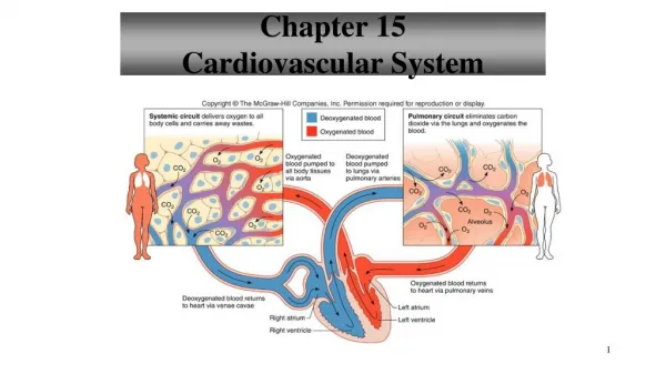
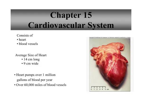
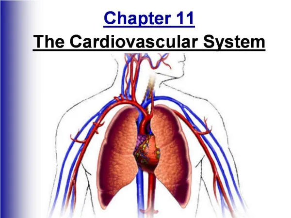
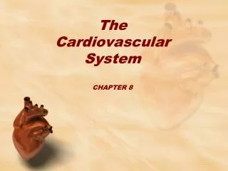
Cardiovascular SystemChapter 13 Objectives: Identify structures and functions of the cardiovascular system. Trace the flow of blood through the body.
Coverings of the Heart • Pericardium: (?) • Visceral (?)pericardium (“epicardium”(?)) • Innermost layer • Covers the heart • Parietal (?)pericardium • Outer layer of visceral pericardium • Forms inner lining of fibrous pericardium • Fibrous (?)pericardium • Dense connective tissue attached to central portion of diaphragm, posterior of sternum, vertebral column, and large blood vessels attached to heart
Pericardial Cavity • Space between parietal and visceral pericardium layers • Filled with serous fluid to reduce friction between the membranes as the heart moves
Wall of the Heart • Epicardium • Outer layer • Corresponds to visceral pericardium • Protects heart by reducing friction • Myocardium • Thick, middle layer • Consists mostly of cardiac muscle tissue that pumps blood out of the heart chambers • Endocardium • Inner layer • Epithelium and connective tissue
Heart Chambers & Valves • ???? • Four hollow chambers: • Upper chambers = atria (atrium) • Lower chambers = ventricles • Atria • Thin walls • Receive blood returning to the heart • Ventricles • Receive blood from the atria • Contract to force blood out of the heart into the arteries
Heart Chambers & Valves, continued….. • Left atrium and ventricle are separated from the right atrium and ventricle by a solid, wall-like septum. • Atrioventricular valve – separates ??? • Tricuspid valve • on the right • “3 cusps” • Bicuspid valve (“mitral valve”) • on the left • “2 cusps” • Allows blood to flow ONLY from atrium to ventricular – no backflow
Heart Chambers & Valves, continued….. • Right atrium receives blood from: • Superior vena cava (???) • Inferior vena cava (???) • Coronary sinus – small vein that drains blood from the myocardium • Right ventricle • Thinner muscular wall than the left ventricle • Only has to pump blood to the lungs • Blood leaves the ventricle through the pulmonary valve, and enters the pulmonary trunk, which divides into the left and right pulmonary arteries.
Heart Chambers & Valves, continued….. • Left atrium receives blood from the lungs through 4 pulmonary veins. • Blood leaves the left ventricle through the aortic valve, into the aorta. • Ventricular wall is thicker – has to pump blood to the rest of the body