Download
1 / 7
70 likes | 95 Views
Explore essential topics on microscopy and staining to understand material in subsequent chapters. Learn about compound light, electron, and fluorescent microscopes. Discover Gram + and Gram - staining procedures, including acid-fast stains and negative staining.

E N D
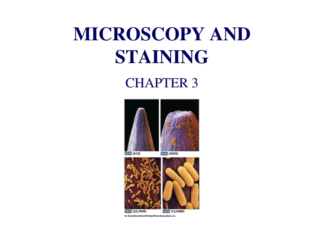
MICROSCOPY AND STAINING CHAPTER 3
CHAPTER 3 MICROSCOPY AND STAINING- Most of the material in this chapter is much better presented in Bio 209 laboratory and the laboratory for Bio 309 where you can actually do hands on work with the microscope and sample preparation. Hence, I will cover only those topics needed to understand material in subsequent chapters. http://www.youtube.com/watch?v=77J4i3uyna0 Compound light microscope for those not taking the laboratory Electron microscope for those not taking The laboratory http://www.youtube.com/watch?v=fToTFjwUc5M Fluorescent microscope for those not taking the laboratory http://www.youtube.com/watch?v=O6JVAUgz0MU
Gram + CV-I complex Gram - CV-I complex is not extracted from Gram positive orgs. Fig. 3.3 Gram stain
This stain produces vivid red color in acid fast organisms such as Mycobacterium leprae, the cause of leprosy or Mycobacterium tuberculosis, the cause of tuberculosis. Fig. 3. 31 The Ziehl-Neelsen acid-fast stain
The clear area around the crystal violet stained organism is the capsule- which does not accept the India ink . Negative staining for capsules reveals a clear area (the capsule, which does not accept stain) in a dark pink background of India ink and crystal violet counter stain Fig. 3.32 Negative staining