HLA and antigen presentation
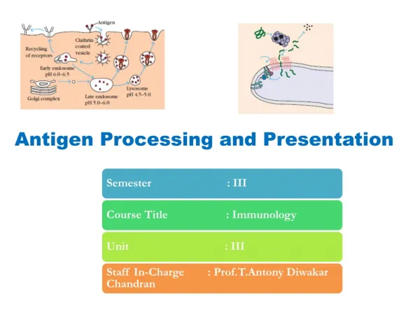
HLA and antigen presentation. Department of Immunology Charles University, 2nd Medical School University Hospital Motol . MHC in adaptive immunity. T cells recognise cell-associated antigens displayed on MHC = major histocompatibility complex. Outline.

HLA and antigen presentation
E N D
Presentation Transcript
HLA and antigen presentation Department of Immunology Charles University, 2nd Medical School University Hospital Motol
MHC in adaptive immunity T cells recognise cell-associated antigens displayed on MHC = major histocompatibility complex
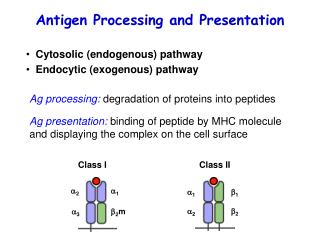
Outline • Adaptive immunity, role of MHC (HLA) • discovery of HLA genes • structure of HLA genes and molecules • polymorphism of HLA molecules • nomenclature of HLA system • HLA association with disease • antigen presentation
HLA – MHC: basic facts • Two groupes of MHC genes: structurally and functionally distinct class I recognition by CD8+ T cells class II recognition by CD4+ T cells • HLA molecules are responsible for the compatibilityof the tissues of genetically different individuals and for the rejection of transplant • MHC genes are codominantly expressed in each individual • monozygotic twins have the same histocompatibility molecules on their cells • MHC genes are the most polymorphic genes present in the genome! (Up to 250 alleles identified for some loci)
MHC expression Class I On all nucleated cells (no MHC on red blood cells, weak expression on cells in CNS) Class II Found on antigen presenting cells
Discovery of Human MHC • Recognition of a graft as self or foreign is an inherited trait • histocompatibility genes: differences between self and foreign were attributed to their genetic polymorphisms • Mouse study: identification of MHC locus • Human MHC In study with transplanted patients discovered „human leukocyte antigens“ HLAs HLA-A, HLA-B, HLA-C (class I MHC genes) In study of mixed leukocyte reaction identified HLA-DR, HLA-DP, HLA-DQ (class II MHC genes
structure of HLA molecules • glycoproteins, heterodimers (two chains) • Structure of HLA molecules of both classes enables antigen binding and contact with T cell receptors. Extracellulary located peptide binding cleft • polymorphic (predominantly in the cleft). • Nonpolymorphic part of the molecule contains binding sites for the T cell molecules CD4 and CD8
HLA class I. molecules 1. Heavy chain α1, α2 domain: polymorphic sites α3 domain: binding of CD8 2. b-2 microglobulin 3. peptide
Structure of HLA class II. molecules 1. α chain α1: polymorphic sites α2: binding of CD4 2. b chain b1: polymorphic sites b2: binding of CD4 3. peptide
HLA and antigens • Most T lymphocytes recognize only peptides • T cells are specific for amino acid sequences of peptides - TCR • Intracellular antigens are presented in connection with HLA class I. - CD8+ T cells recognition • Extracellular antigens are presented in connection with HLA class II. – CD4+ T cells recognition Experiment: • T cell response – only when peptide is attached to APC
HLA and peptides • antigenic peptides in the binding sites of HLA molecules • One MHC - many peptides sharing structural features can bind • Interaction has a very slow on- and off-rate (very stable) • class I. • class II.
HLA genes MHC locus • On chromosome 6 • HLA class III. are soluble molecules as complement, TNF, HSP • Many other proteins involved in antigen presentation
HLA polymorphism • most polymorhic structures from all known systems. • human study: 1000 donors, HLA-A, B genotyping • Over half the group had a combination that was unique. • Another 111 donors had a set of these molecules that they shared with only one other person in the group. • The most frequent phenotype (HLA-A1, HLA-A3, HLA-B7, and HLA-B8) was found in 11 donors.
HLA polymorphism – why? Pathogen driven mechanisms Pathogens tend to escape Heterozygotes have advantage Frequency-dependent selection: the individual with the rarest allele has the best chance to survive an infection cheetahs (low polymorphism): extremly susceptible to infectious diseases vertebrate species can detect MHC genotype by smell!
HLA haplotypes in a typical family • Haplotype is combination of allelic forms of HLA molecules on one chromosome. • We inherit 3 types of heavy chains for HLA class I. molecules from each parent . • Everybody expresses 6 different types of HLA class I. molecules unless honmozygous status for some of the types was inherited.
MHC control transplantat survival • A graft is compatible only if there is a complete match at all MHC alleles, i.e. a two haplotype match for all MHC loci
lysozom APC ER, Golgi HLA class II. antigen TCR CD4 T lymfocyte Function of MHC • Recognition of antigen by T cells is necessary for induction of the immune response. • exogenous antigen presentation
Antigen presenting cells Other population of APC • Vascular endothelial cells: function as APC is inducible • Various epithelial and mesenchymal cells: inducible Most important population of APC
Dendritic cells in antigen presentation Dendritic cells - most effective population in T cell activation - used as immunoterapeutic tools in cancer vaccines Immature DC: capture antigens in periphery Mature DC: activation of T lymphocytes in lymphatic nodes
Antigen Presentation and Dendritic Cells Immature (a), maturing (b), and mature (c) dendritic cells stained for MHC class II (green) and lysosomal marker Lamp-1 (red). From Mellman et al., TICB 8: 231 (1998) J. R. Lingappa, Pabio 552
Exogenous antigens • Exogenous antigens (inhaled, ingested, or injected) are taken up by "professional" antigen-presenting cells • These include: phagocytic cells like macrophages and dendritic cells B lymphocytes which are responsible for producing antibodies against the antigen. • All these cells express HLA class II. molecules
Exogenous antigen processing • Invariant Chain (Ii)-Contributes to: • proper folding of the alpha and beta chains in the ER • preventing in advertent association with endogenous peptides • directing immature MHC II through the secretory pathway CLIP (Class II-associated invariant chain peptide)-Degradation product derived from the Invariant chain. HLA-DM Responsible for displacing CLIP and allowing peptide to bind once MHC has reached the endosome. HLA-DO Negative regulator of HLA-DM activity
TAP (transporter associated with antigen presentation) • Transport associated protein - TAP is responsible for the peptide transport from cytoplasm to ER. • Proteins are degraded to peptide in proteasome. • The peptides are picked up by TAP proteins and transported from the cytosol into the RER where they assemble with • the transmembrane polypeptide and beta-2 microglobulin. • this trimolecular complex then moves through the Golgi apparatus and is inserted in the plasma membrane
Imunodeficiences - MHC defect • Bare lymphocyte syndrome: mutation in genes regulating class II MHC transcription - reduced number of CD4+ T cells in periphery - defective activation of CD4+ T cells - fatal, treatment: BM transplantation • Class I MHC deficiencies: - decreased number of CD4+ T cells in periphery - caused by TAP1, TAP2 - patients suffer from respiratory tract bacterial infection
HLA-associated diseases HLAPatientsControls Ankylosing spondylitis B27 90% 9% Type 1 diabetes DR352% 23% DR4 74% 24% DR3 or DR4 93% 43% Multiple sclerosis DR2 86% 33% Rheumatoid arthritis DR4 81% 24% NarcolepsyDR2 >95% 33%
lysozome APC APC ER, Golgi ER, Golgi B lymfocyte HLA class I. antigen TCR HLA class II. antigen TCR HLA class II. antigen TCR CD8 CD4 CD4 T lymfocyte T lymfocyte T lymfocyte Summary -antigen presentation pathways endogenous exogenous B lymfocytes cell destruction immune response antibody production