Download

1 / 54
590 likes | 1.06k Views
DEPARTMENT OF IMMUNOLOGY. Antigen Presenting Cell and Antigen Presentation. Haifeng Gao hfgao@fudan.edu.cn. Antigen Presenting Cells Antigen Processing Antigen Presentation. First line of defense Native Non-specific No memory to antigens. Innate Immunity. Acquired Specific

E N D
DEPARTMENT OF IMMUNOLOGY Antigen Presenting Cell and Antigen Presentation Haifeng Gao hfgao@fudan.edu.cn
Antigen Presenting Cells Antigen Processing Antigen Presentation
First line of defense Native Non-specific No memory to antigens Innate Immunity Acquired Specific Memory to antigens Recognition by receptor Adaptive Immunity
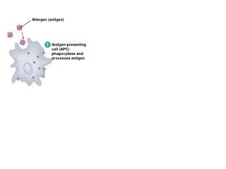
APC Antigen fragment MHC TCR Antigen Presenting Cell, APC Pathogens T Cell
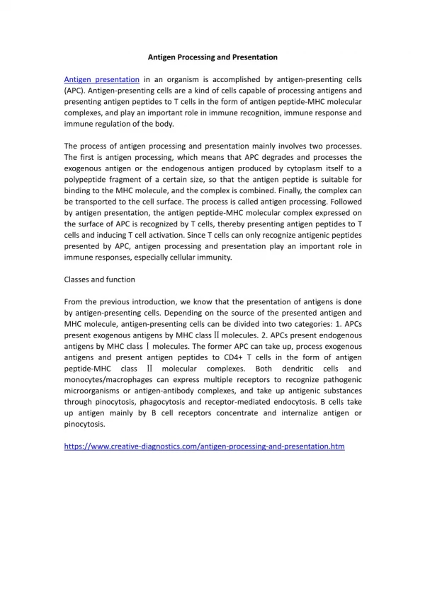
CONCEPT APCs are immunocytes that can uptake, process and present antigens to other lymphocytes.
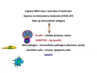
CLASIFICATION Professional APC: constitutively express MHC II molecules MHC I
Professional APCs Dendritic Cells (DCs) Macrophages (M) B Lymphocytes
Non-professional APCs Endothelial Cells Epithelial Cells Fibroblast Cells …… Non-professional APC: express MHC I molecules
I. Dendritic Cells (DCs) Ralph.M.Steinman, 1973
Migration maturation Immature DC Mature DC
Immature DC Strong antigen uptaking and processing function Mature DC High level of MHC molecules and co-stimulatory molecules
Fluorescent Microscopy DCs in peripheral tissues DCs in lymphoid tissues
Scanning Electron Microscopy DC in peripheral tissues DC in lymphoid tissues
CHARICTERISTICS OF DC effectively uptake antigen effectively present antigen Express high levels of MHC II and co-stimulatory molecules (such as B7) effectively prime naïve T cells
Classification • LC: Langekhan's cell • IDC: Interdigitating DC • FDC: Follicular DC
Function Immune Regulation Antigen Presentation Immune Tolerance
Mononuclear Phagocyte System,MPS Bone marrow pluripotent stem cells Mononuclear cells Macrophages
Macrophages Phagocytosis and antigen presentation
Function Phagocytosis Antigen Presentation Cytotoxicity Wound Healing Secret Cytokines
III. B Lymphocytes BCR B Cell Receptor (BCR): binding with specific antigen
Summary APCs are immunocytes that can uptake, process and present antigens to lymphocytes. DCs are the most powerful APC in vivo, which can effectively stimulate naïve T cells to proliferate. M and B cells can only stimulate activated or memory T cells.
? Next Chapter: How APC processing the antigen
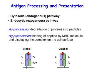
1、MHC class I molecule way APC:Nucleated cells Antigen:Endogenous peptides Molecule:MHC class I Processed area:Proteasome (polyubiquitination) Combined area:ER
Nobel Prize in Chemistry for 2004 “for the discovery of ubiquitin-mediated protein degradation" Aaron Ciechanover Avram Hershko Irwin Rose ubiquitin / proteasome system
Endogenous peptides Proteasomes are cytoplasmic organelles that degrade cytoplasmic proteins 6-30 Amino acids
Hydrophobic transmembrane domain Lumen of ER Lumen of ER Peptide Peptide Peptide Peptide Peptide Peptide Peptide Peptide Peptide Peptide Peptide ER membrane ER membrane TAP-1 TAP-1 TAP-1 TAP-1 TAP-1 TAP-1 TAP-1 TAP-1 TAP-1 TAP-1 TAP-1 TAP-2 TAP-2 TAP-2 TAP-2 TAP-2 TAP-2 TAP-2 TAP-2 TAP-2 TAP-2 TAP-2 Cytosol Cytosol ATP-binding cassette (ABC) domain Peptide antigens from proteasome Transporter has preference for 8~12 amino acid peptides with hydrophobic C termini.
Peptide Peptide Peptide Peptide Peptide Peptide Peptide Peptide Peptide Peptide Peptide TAP-1 TAP-1 TAP-1 TAP-1 TAP-1 TAP-1 TAP-2 TAP-2 TAP-2 TAP-2 TAP-2 TAP-2 TAP-2 TAP-2 TAP-2 TAP-2 TAP-2 TAP-1 TAP-1 TAP-1 TAP-1 TAP-1 Endoplasmic reticulum Maturation and loading of MHC class I B2-M binds and stabilises floppy MHC Tapasin, calreticulin, TAP 1 & 2 form a complex with the floppy MHC Calnexin binds to nascent class Ia chain until b2-M binds Cytoplasmic peptides are loaded onto the MHC molecule and the structure becomes compact
2、MHC class II molecule way APC:Professional APC Antigen:Exogenous peptides Molecule:MHC class II Processed area:Endosome Combined area:M II C
(1)Exogenous peptides Endosome Cathepsin 、Catalase 10~30 Amino acids
(2)MHC class II molecule Ia-associated invariant chain, Ii (Ii)3 nine polymer MHC class II compartment, M II C Ii chain degraded
Function of Ii chain Promote MHC class II molecule to assemble Prevent MHC class II molecule to combine with other peptides Promote MHC class II molecule transportation
The invariant chain is cleaved to leave a peptide fragment, CLIP, bound to the MHC class II molecule CLIP (class II-associatedinvariant-chain peptide)
Cell surface Endosomes Uptake Class II associated invariant chain peptide (CLIP) (abIi)3 complexes directed towards endosomes by invariant chain Cathepsin L degrades Invariant chain CLIP blocks groove in MHC molecule MHC Class II containing vesicles fuse with antigen containing vesicles