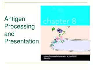
Antigen Processing and Presentation
Antigen Processing and Presentation. Processing of Antigen Is Required for Recognition by T Cells. Most Cells Can Present Antigen with Class I MHC; Presentation with Class II MHC Is Restricted to APCs.

Antigen Processing and Presentation
E N D
Presentation Transcript
Processing of Antigen Is Required for Recognition by T Cells
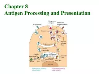
Most Cells Can Present Antigen with Class I MHC; Presentation with Class II MHC Is Restricted to APCs 1)Dendritic cells are the most effective of the antigen presenting cells. Because these cells constitutively express a high level of class II MHC molecules and costimulatory activity, they can activate naive TH cells.
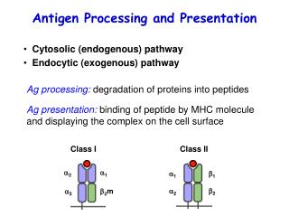
Macrophages must be activated by phagocytosis of particulate antigens before they express class II MHC molecules or the co-stimulatory B7 membrane molecule. • B cells constitutively express class II MHC molecules but must be activated before they express the co-stimulatory B7 molecule. Endogenous Antigens: The Cytosolic Pathway Peptides for Presentation Are Generated by Protease Complexes Called Proteasomes
Peptides Are Transported from the Cytosol to the Rough Endoplasmic Reticulum The proteasomes involved in antigen processing include two subunits encoded within the MHC gene cluster, LMP2 and LMP7, and a third non-MHC protein, LMP10 (also called MECL-1). All three are induced by increased levels of the T-cell cytokine IFN gamma.
Peptides Assemble with Class I MHC Aided by Chaperone Molecules
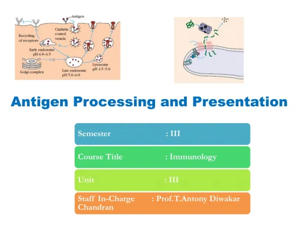
Exogenous Antigens: The Endocytic Pathway Peptides Are Generated from Internalized Molecules in Endocytic Vesicles The endocytic pathway appears to involve three increasingly acidic compartments: early endosomes (pH 6.0–6.5); late endosomes, or endolysosomes (pH 5.0–6.0); and lysosomes (pH 4.5–5.0). Internalized antigen moves from early to late endosomes and finally to lysosomes, encountering hydrolytic enzymes and a lower pH in each compartment. Lysosomes, for example, contain a unique collection of more than 40 acid-dependent hydrolases, including proteases, nucleases, glycosidases, lipases, phospholipases, and phosphatases. Within the compartments of the endocytic pathway, antigen is degraded into oligopeptides of about 13– 18 residues,which bind to class II MHC molecules.
The Invariant Chain Guides Transport of Class II MHC Molecules to Endocytic Vesicles
Peptides Assemble with Class II MHC Molecules by Displacing CLIP