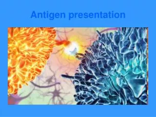
Antigen-presenting cells and antigen presentation
500 likes | 1.3k Views
Antigen-presenting cells and antigen presentation. 1.Nature of antigen-presenting cells 2. Antigen processing and presentation. Antigen Presenting Cell (APC). Section 1 Nature of antigen-presenting cells.

Antigen-presenting cells and antigen presentation
E N D
Presentation Transcript
Antigen-presenting cells and antigen presentation 1.Nature of antigen-presenting cells 2. Antigen processing and presentation
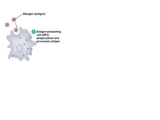
Section 1 Nature of antigen-presenting cells Antigen-presenting cells (Antigen Presenting Cell, APC) are the immunocytes that can uptake, process and present antigens to lymphocytes.
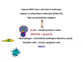
Professional APC Dendritic cells, mononuclear phagocyte system, B cells, and APC that constitutively express MHC II molecules Non-professional APC
DC are the most powerful APC in vivo, which can effectively stimulate naïve T cells to proliferate. Yet M and B cells can only stimulate activated or memory T cells.
Multiple hematopoietic stem cell Lymphoid stem cell Myeloid stem cell Neutrophil Myeloid DC Monocyte NK cell Lymphoid DC T cell B cell Macrophage Myeloid DC Origination of dendritic cells(DC)
Tissue distribution : Lymphoid DC:interdigitating DC, marginal DC Non-lymphoid DC: interstitial DC, LC, gastrointestinal epithelial DC Humoral DC: veiled cell Blood DC
Differentiation and development of DC • precusor • immature stage (immature DC) • migratory stage • mature stage(mature DC) Immature DC Distribution: many solid organs and non lymphatic epithelia Major role: uptake and process antigen Normally, most DC are immature DC.
Mature DC Distribution: T lymphocyte areas in lymph node, spleen, and Peyer’s node, blood Characteristics: abundantly express MHC II、MHC I、CD80、CD86、CD40、ICAM-1、HSP、CD1a、CD11c、CD83 Major role: antigen presentation, present peptide:MHC complex to T cells
Antigen uptaking pathways by DC • macropinocytosis • receptor-mediated endocytosis • phagocytosis
2. Mononuclear macrophages M: Origination and distribution Bone marrow pluripotent stem cells-mononuclear cells-macrophages
Antigen processing and presentation of M • endocytosis or internalization: • phagocytosis • Pinocytosis • receptor-mediated endocytosis Only those M in vigorous metabolism have the capacity of antigen presentation.
Macrophagocytosis mIg mediated endocytosis Complement receptor mediated endocytosis Pinocytosis Fc receptor mediated endocytosis Uptake of exogenous antigens
3 B lymphocytes Receptor-mediated endocytosis Non-specific pinocytosis
Dendritic cell macrophage B cell Staining of APC
Section 2 Antigen processing and presentation 1 Antigen uptaking
DC capture antigens : Macropinocytosis Receptor-mediated endocytosis Phagocytosis
Mononuclear macrophage capture antigens: Phagocytosis Pinocytosis Receptor-mediated endocytosis
B cells captureantigens: Non-specific pinocytosis Antigen-specific receptor mediated
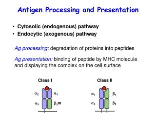
2. Antigen processing 1、Endogenous pathway Antigens: intracellular synthesized antigens Presenting molecules:MHC-I Processing region: Proteosome (polyubiquitination) Binding region: ER
Cell surface cytoplasm Golgi complex Endoplasmic reticulum calnexin MHC Ia chain Degraded antigens proteasome Endogenous antigens Processing and presentation of endogenous antigens
(1) Production of endogenous antigenic peptide Ubiquitination of endogenous antigens Degradation of endogenous antigens in proteosome Peptides containing 8-12 amino acidresidues
(2) Transportation of endogenous antigenic peptide Endogenous antigenic peptide enter endoplasmic reticulum: Transporter associated with antigen processing(TAP)
(3) MHC class I molecules peptide loading Class I molecules associate with chaperon: Bind to endogenous antigenic peptides Enter Golgi complex to undergo glycosylation modification Traffick to cell surface in exocytic vesicles and recognized by CD8+ T cells
Membrane Antigen-MHC class 1 molecule complex β2 microglobulin Heavy chain of antigen-MHC class 1 molecule Golgi complex β2 microglobulin Calnexin Antigenic peptide Endogenous antigen Presentation of endogenous antigens Proteasome
2. Presenting pathway of exogenous antigens(Exogenous pathway) Antigen: extracellular antigens Presenting molecules: MHC-II Processing region: endosome Binding region: MIIC、CIIV
Cell surface Exogenous antigens Fusion Early endosome Late endosome Antigen degradation Golgi complex MHC class 2 molecule Ii chain Endoplasmic reticulum Processing and presentation of exogenous antigens
Antigen processing compartment Endosome MHC class II、HLA-DM, and exogenous antigenic peptide are found in abundance.
(2)Degradation of protein antigen Peptides containing 12-20 amino acid residues Endopeptidase Exopeptidase
MHC class II molecules are transported from ER to endosome. Calnexin Ia associated invariant chain(Ii chain): Class II associated invariant chain peptide (CLIP) Form nonamer with , chain Enter endosome through trans-Golgi network
Function of Ii To prevent degradation of MHC class II molecule during its transportation CLIP binds to the grooves in MHC class II molecule
(4)Ii degradation in endosome (5)Class II molecules peptide loading
(6)Presentation of exogenous antigens Exocytic vesicles Expressed on APC Recognized by CD4+ T cells
Antigen Antigen Antigenic peptide-MHC class 2 molecule complex internalization Phagocytosis B cell Macrophage Endosome/lysosome Golgi complex ER Antigenic peptide-MHC class 2 molecule complex αβIi complex Ii chain Presentation of exogenous antigens
Comparison of endogenous and exogenous antigen-presenting pathway Characteristics Endogenous pathway exogenous pathway Presenting molecule MHC-I MHC-II Responding T cells CD8+ T cells CD4+ T cells Antigen resources endogenously synthesized exogenous uptaken Synthesizing region of antigen peptides proteosome endosome Chaperon Calnexin, TAP, tapasin Calnexin, Ii chain Presenting cells all nucleated cells professional APC
3. Antigen presentation 1. Basic procedure of antigen presentation
CD8+ T cells Tumor cells
2.Cross presentation of antigens by MHC molecules (Cross priming pathway)
Master cell types, definition and characteristics of APC • Master types of professional APC • Master the pathway and procedure of exogenous and endogenous antigen presentation • Familiarize antigen cross presentation • Understand the distribution and characteristics of various kinds of professional APC • Understand development, differentiation, maturation and migration of DC