Download
1 / 75
750 likes | 773 Views
This article provides an overview of the digestive system, including its functions and the organs involved. Topics covered include digestion, absorption of nutrients, metabolism, and the roles of the alimentary canal and accessory digestive organs. The anatomy and functions of the mouth, pharynx, esophagus, stomach, small intestine, and large intestine are also discussed.

E N D
Basics • Digestion • Breakdown of ingested food • Absorption of nutrients into the blood • Metabolism • Production of cellular energy (ATP) • Constructive and degradative cellular activities
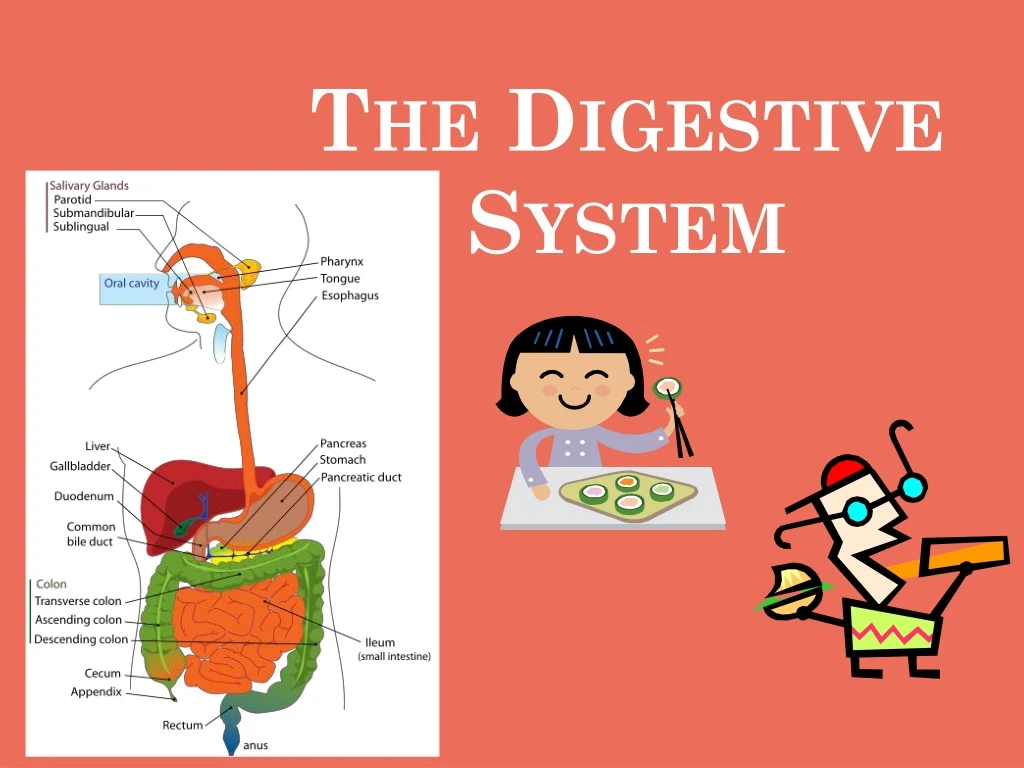
Digestive Organs • Two main groups • Alimentary canal – continuous coiled hollow, muscular tube (aka gastrointestinal [GI] tract) open at both ends • Performs all digestive functions • Ingest, digest, absorb, defecate • Accessory digestive organs • Assist process of digestion
If it’s where food goes through, it’s the GI tract • If it’s attached to GI tract, it’s an accessory organ Figure 14.1
1. Mouth (Oral Cavity) • Mechanical breakdown • Food is physically broken down by mastication (chewing) • Chemical digestion • Food is mixed with saliva which breaking of starch into maltose by enzyme salivary amylase • Initiation of deglutition (swallowing ) by the tongue • Allowing for the sense of taste • 5 tastes: • Sweet Sour Salty Bitter Umami (savory/tasty)
Anatomy of Mouth • Lips (labia) • Cheeks • Hard palate – forms the anterior roof • Soft palate – forms the posterior roof • Uvula – fleshy projection of the soft palate; no function • Vestibule – space between lips and teeth and gums Figure 14.2a
Oral cavity – behind the teeth • Tongue – attached at hyoid and styloid, and by lingual frenulum • Tonsils • Palatine tonsils • Lingual tonsil • Part of lymphatic (immune) system Figure 14.2a
2. Pharynx • Serves as a passageway for air and food • Food movement via alternating contractions of the muscle layers (peristalsis)
Pharynx Anatomy • Nasopharynx – not part of the digestive system; up to the nose • Oropharynx – posterior to oral cavity; back of throat • Laryngopharynx – below the oropharynx and connected to the esophagus
3. Esophagus • Runs from pharynx to stomach through the diaphragm • Conducts food by peristalsis (slow rhythmic squeezing) • Passageway for food only (respiratory system branches off after the pharynx) • Timeline: 5-8 seconds!
4. Stomach • Acts as a storage tank for food • Can hold 4 L (1 gal) of food!! • Site of physical food breakdown • Chemical breakdown of protein begins • Lined with columnar epithelium with … • Mucus neck cells– mucus • Chief cells – pepsinogen (digest protein) • Parietal cells – HCl (hydrochloric acid, pH~1.5) • Enteroendocrine cells – gastrin • Gastric cells – gastrin juice • Delivers chyme (processed food) to the small intestine
DO NOT COPY Pepsi & Pepsin • Caleb Bradham of North Carolina was a pharmacist. • His most popular beverage was something he called "Brad's drink" made of carbonated water, sugar, vanilla, rare oils, pepsin and kola nuts. • He later on renamed his beverage Pepsi Cola and it advertised it as “Exhilarating, invigorating, aids digestion.”
Stomach Anatomy • Located on the left side of the abdominal cavity • Food enters at the cardioesophageal sphincter – circular muscle that acts as “gatekeeper” • Food empties into the small intestine at the pyloric sphincter • Timeline: 2-6 hours • Contains rugae – internal folds of the mucosa • Layers of peritoneum attached to the stomach • Lesser omentum – attaches the liver to the lesser curvature • Greater omentum – attaches the greater curvature to the posterior body wall • Contains fat to insulate, cushion, and protect abdominal organs (see Dr. Oz on Oprah)
Structure of the Stomach Mucosa Figure 14.4b–c
5. Small Intestine • body’s major digestive organ – averages 7m (>23 feet) long! • Site of nutrient absorption into the blood • Timeline: 3-5 hours • Muscular tube extending from the pyloric sphincter to the ileocecal valve • Suspended from the posterior abdominal wall by the mesentery • Double-layer of peritoneum
Divided up into three sections: • Duodenum • Attached to the stomach • Curves around the head of the pancreas • Jejunum • Attaches anteriorly to the duodenum • Ileum • Extends from jejunum to large intestine
Many chemicals involved in digestion in small intestine • Enzymes from intestinal cells • Enzymes from pancreas • Bile from gall bladder
Absorption is done through many villi • Fingerlike structures formed by the mucosa • Give the small intestine more surface area • Microvilli on absorptive cells of villi add for super absorption • Small projections of the plasma membrane • Found on absorptive cells
Absorption into bloodstream carried out by • Absorptive cells • Blood capillaries • Lacteals (specialized lymphatic capillaries) Figure 14.7b
6. Large Intestine • Larger in diameter, but shorter than the small intestine – 1.5 m long • Frames the internal abdomen • Absorption of water • Eliminates indigestible food from the body as feces • Does NOT participate in digestion of food • Goblet cells produce mucus to act as a lubricant
Divided up into • Colon • Ascending – up • Transverse – across • Descending – down • S-shaped sigmoidal • Rectum • Anal canal • Cecum – saclike projection, hangs from first part • Appendix – twisted section that often traps bacteria & gets infected (appendicitis) • Timeline: 4-72 hours!
7. Anus • Ending of anal canal • Contains two sphincters which work to control passage of fecal matter • Internal involuntary sphincter • Signals us that it’s time to expel feces • External voluntary sphincter • Control opening of sphincter until ready
Accessory Digestive Organs • Salivary glands • Teeth • Pancreas • Liver • Gall bladder
Salivary Glands • Saliva-producing glands • Parotid glands – located anterior to ears • Submandibular glands - under mandible • Sublingual glands – under tongue
Produce saliva • Mixture of mucus and serous fluids • Helps to form a food bolus (ball of masticated food) • Contains salivary amylase to begin starch digestion • Dissolves chemicals so they can be tasted
2. Teeth • role is to masticate (chew) food • Humans have two sets of teeth • Deciduous (baby or milk) teeth • 20 teeth fully formed by age two • Permanent teeth • Replace deciduous teeth beginning ages of 6 to 12 • full set is 32 teeth, some people do not have wisdom teeth
3. Pancreas • Produces a wide spectrum of digestive enzymes that break down all categories of food • Enzymes secreted into duodenum • Alkaline fluid introduced with enzymes neutralizes acidic chyme • Endocrine products of pancreas: • Insulin • Glucagon
4. Liver • Largest gland in the body • Located on the right side of the body under the diaphragm • Consists of four lobes suspended from the diaphragm and abdominal wall by the falciform ligament • Connected to the gall bladder via the common hepatic duct
Produces bile – highly bitter green liquid containing: • Bile salts • Bile pigment (mostly green bilirubin from the breakdown of hemoglobin) • Cholesterol • Phospholipids • Electrolytes
5. Gall Bladder • Sac found in hollow fossa of liver • Stores bile from the liver by way of the cystic duct • Bile is introduced into the duodenum in the presence of fatty food • Gallstones can cause blockages
B. Six Processes of Digestive System • Ingestion – getting food into the mouth • Mechanical Digestion – physically breaking food down • Propulsion – moving foods from one region of the digestive system to another • Chemical Digestion – chemically breaking down food • Absorption – getting nutrients/water into blood stream • Defecation – expelling wastes
Ingestion • Voluntary process of getting food into mouth • Mechanical Digestion • Mixing of food in the mouth by the tongue • Churning of food in the stomach • Segmentation in the small intestine • Movement of food back & forth serving to mix it with digestive juices
Propulsion • Food is processed by more than one digestive organ so must be propelled from one to another. Done via peristalsis • Peristalsis – involuntary action where alternating waves of contraction & relaxation of smooth muscles squeeze food along GI tract
Food must first be well mixed • Rippling peristalsis occurs in lower stomach • pylorus meters out chyme into the small intestine (30 ml at a time) • stomach empties in four to six hours Figure 14.15
Chemical Digestion • Enzymes break down food molecules into their building blocks • Each major food group uses different enzymes • Carbohydrates simple sugars • Proteins amino acids • Fats fatty acids and alcohols • Absorption • End products of digestion absorbed in blood or lymph • Food must enter mucosal cells, then into blood or lymph capillaries • Defecation • Elimination of indigestible substances as feces
C. Control of Digestive Activity • Mostly controlled by reflexes • Chemical and mechanical receptors are located in organ walls that trigger reflexes • Stimuli include: • Stretch of the organ • pH of the contents • Presence of breakdown products • Reflexes include: • Activation or inhibition of glandular secretions • Smooth muscle activity
D. Activities by GI Organs • Mouth-Esophagus (Deglutition = Swallowing) • Buccal phase • Voluntary; occurs in mouth; food formed into bolus • Bolus forced into pharynx by tongue • Pharyngeal-esophageal phase • Involuntary transport of bolus • All passageways blocked • Tongue blocks off mouth • Soft palate (uvula) blocks nasopharynx • Epiglottis blocks larynx • Cardioesophageal sphincter opened when bolus presses against it
Deglutition (Swallowing) Figure 14.14
Stomach • Gastric juice regulated by neural and hormonal factors • Presence of food and/or falling pH (due to HCl) causes release of gastrin • Gastrin causes stomach glands to produce protein-digesting enzymes pepsin & rennin • HCl activates pepsinogen to become pepsin for protein digestion • HCl provides hostile environment for microorganisms (except Helicobacter pylori) • Only absorption occurring stomach is alcohol and aspirin
Small intestine • Enzymes from brush border break double sugars (lactose) into simple sugars (galactose & glucose) • Complete some protein digestion • Pancreatic enzymes play the major digestive function • Help complete digestion of starch (pancreatic amylase) • Carry out about half of all protein digestion (trypsin, etc.) • Responsible for fat digestion (lipase) • Digest nucleic acids (nucleases) • Alkaline content neutralizes acidic chyme
Release of pancreatic juice stimulated by: • Vagus nerve • Local hormones • Secretin: causes liver to produce more bile; pancreas to release more alkaline juice • Cholecystokinin (CCK): stimulates gall bladder to release more bile; pancreas to release more enzymes
Large Intestine • No digestive enzymes are produced • Resident bacteria digest remaining nutrients (Escherichia coli) • Produce some vitamin K and B • Release gases • Water and vitamins K and B are absorbed • Remaining materials are eliminated via feces
Sluggish peristalsis • Mass movements • Slow, powerful movements • Occur three to four times per day • Presence of feces in the rectum causes a defecation reflex • Internal anal sphincter is relaxed • Defecation occurs with relaxation of the voluntary (external) anal sphincter