Download
1 / 37
370 likes | 545 Views
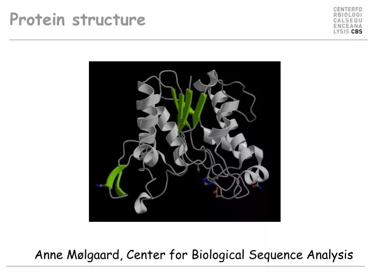
Protein structure. Anne Mølgaard, Center for Biological Sequence Analysis. “Could the search for ultimate truth really have revealed so hideous and visceral-looking an object?”. Max Perutz, 1964 on protein structure. John Kendrew, 1959 with myoglobin model.

E N D
Protein structure Anne Mølgaard, Center for Biological Sequence Analysis
“Could the search for ultimate truth really have revealed so hideous and visceral-looking an object?” Max Perutz, 1964 on protein structure John Kendrew, 1959 with myoglobin model
Holdings of the Protein Data Bank (PDB): Sep. 2001 Feb. 2005 X-ray 13116 25350 NMR 2451 4383 theoretical 338 0 total 15905 29733
Methods for structure determination • X-ray crystallography • Nuclear Magnetic Resonance (NMR) • Modeling techniques
Modeling Only applicable to ~50% of sequences Fast Accuracy poor for low sequence id. • There is still need for experimental structure determination!
Structual genomics consortium (SGC) • The SGC deposited its 275th structure into the Protein Data Bank in August 2006 • currently operating at a pace of 170 structures per year • at a cost of USD$125,000 per structure. • Scientific highlights include: • several (> 1!!) novel structures of protein kinases • completing the structural descriptions of the human 14-3-3 • adenylate kinase and cytosolic sulfotransferase protein families • human chromatin modifying enzymes; human inositol phosphate signaling • and a significant number of structures from human parasites.
Amino acids http://www.ch.cam.ac.uk/magnus/molecules/amino/
Amino acids A – Ala C – Cys D – Asp E – Glu F – Phe G – Gly H – His I – Ile K – Lys L – Leu M – Met N – Asn P – Pro Q – Gln R – Arg S – Ser T – Thr V – Val W – Trp Y - Tyr Livingstone & Barton, CABIOS, 9, 745-756, 1993
Levels of protein structure • Primary • Secondary • Tertiary • Quarternary
Primary structure MKTAALAPLFFLPSALATTVYLA GDSTMAKNGGGSGTNGWGEYL ASYLSATVVNDAVAGRSAR…(etc)
Ramachandran plot left-handed -helix -sheet -helix
Hydrophobic core • Hydrophobic side chains go into the core of the molecule – but the main chain is highly polar • The polar groups (C=O and NH) are neutralized through formation of H-bonds
Secondary structure -helix C=O(n)…HN(n+4) -sheet (anti-parallel)
… and all the rest • 310 helices (C=O(n)…HN(n+3)), p-helices (C=O(n)…HN(n+5)) • b-turns and loops (in old textbooks sometimes referred to as random coil)
- N C + The -helix has a dipole moment
Two types of -sheet: parallel anti-parallel
Tertiary structure (domains, modules) Rhamnogalacturonan acetylesterase (1k7c) Rhamnogalacturonan lyase (1nkg)
Quaternary structure B.caldolyticus UPRTase (1i5e) B.subtilis PRPP synthase (1dkr)
Classification schemes • SCOP • Manual classification (A. Murzin) • CATH • Semi manual classification (C. Orengo) • FSSP • Automatic classification (L. Holm)
Levels in SCOP Class # Folds # Superfamilies # Families • All alpha proteins 202 342 550 • All beta proteins 141 280 529 • Alpha and beta proteins (a/b) 130 213 593 • Alpha and beta proteins (a+b) 260 386 650 • Multi-domain proteins 40 40 55 • Membrane and cell surface • proteins 42 82 91 • Small proteins 72 104 162 • Total 887 1447 2630 http://scop.berkeley.edu/count.html#scop-1.67
Major classes in SCOP • Classes • All alpha proteins • Alpha and beta proteins (a/b) • Alpha and beta proteins (a+b) • Multi-domain proteins • Membrane and cell surface proteins • Small proteins
Folds* • Proteins which have >~50% of their secondary structure elements arranged the in the same order in the protein chain and in three dimensions are classified as having the same fold • No evolutionary relation between proteins • *confusingly also called fold classes
Superfamilies • Proteins which are (remote) evolutionarily related • Sequence similarity low • Share function • Share special structural features • Relationships between members of a superfamily may not be readily recognizable from the sequence alone
Families • Proteins whose evolutionarily relationship is readily recognizable from the sequence (>~25% sequence identity) • Families are further subdivided into Proteins • Proteins are divided into Species • The same protein may be found in several species
Links • PDB (protein structure database) • www.rcsb.org/pdb/ • SCOP (protein classification database) • scop.berkeley.edu • CATH (protein classification database) • www.biochem.ucl.ac.uk/bsm/cath • FSSP (protein classification database) • www.ebi.ac.uk/dali/fssp/fssp.html
Why are protein structures so interesting? They provide a detailed picture of interesting biological features, such as active site, substrate specificity, allosteric regulation etc. They aid in rational drug design and protein engineering They can elucidate evolutionary relationships undetectable by sequence comparisons
NH2 Asp His Ser COOH Inferring biological features from the structure 1deo Topological switchpoint
Inferring biological features from the structure Active site Triose phosephate isomerase (1ag1) (Verlinde et al. (1991) Eur.J.Biochem. 198, 53)
Engineering thermostability in serpins • Overpacking • Buried polar groups • Cavities Im, Ryu & Yu (2004) Engineering thermostability in serine protease inhibitors PEDS, 17, 325-331.
Evolution... Structure is conserved longer than both sequenceandfunction
Rhamnogalacturonan acetylesterase (A. aculeatus) (1k7c) Serine esterase (S. scabies) (1esc) Platelet activating factor acetylhydrolase (Bos Taurus) (1wab)
Rhamnogalacturonan acetylesterase Serine esterase Platelet activating factor acetylhydrolase Mølgaard, Kauppinen & Larsen (2000) Structure, 8, 373-383.
"We wish to suggest a structure for the salt of deoxyribose nucleic acid (D.N.A.). This structure has novel features which are of considerable biological interest…. • …It has not escaped our notice that the specific pairing we have postulated immediately suggests a possible copying mechanism for the genetic material." • J.D. Watson & F.H.C. Crick (1953) Nature, 171, 737.