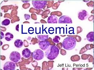
Leukemia
Leukemia. DR. SHAHID MAHMOOD. Leukemia. Is a malignant hematological disorder characterized by a proliferation of abnormal white cells that infiltrate the bone marrow, peripheral blood and organs 4 main types of leukemia Acute or chronic Myelogenous or Lymphocytic . FAB CLASSIFICATION.

Leukemia
E N D
Presentation Transcript
Leukemia DR. SHAHID MAHMOOD RTT 341- Leukemia
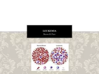
Leukemia • Is a malignant hematological disorder characterized by a proliferation of abnormal white cells that infiltrate the bone marrow, peripheral blood and organs • 4 main types of leukemia • Acute or chronic • Myelogenous or Lymphocytic RTT 341- Leukemia
FAB CLASSIFICATION • M0: minimally differentiated • M1: myeloblastic leukemia without maturation • M2: myeloblastic leukemia with maturation • M3: hypergranular promyelocytic leukemia • M4: myelomonocytic leukemiaM4Eo: variant, increase in marrow eosinophils • M5: monocytic leukemia • M6: erythroleukemia (DiGuglielmo's disease) • M7: megakaryoblastic leukemia RTT 341- Leukemia
Types of Leukemia Acute Leukemia • progresses quickly • characterized by the proliferation of undifferentiated cells in the bone marrow • Chronic Leukemia • slower progression • uncontrolled expansion of mature cells RTT 341- Leukemia
Types of Leukemia • Acute and chronic leukemias are further subdivided into: • Myelogenous Leukemias • those that are from hemopoietic stem cells • Lymphocytic Leukemias • arise from any other cells in the bone marrow RTT 341- Leukemia
Four main subtypes of leukemia: • The four subtypes account for 50% of all leukemia in the U.S • 1. ALL: Acute Lymphocytic Leukemia • 2. AML: Acute Myelogenous Leukemia • is referred to as ANLL- acute nonlymphocytic leukemia • 3. CLL: Chronic Lymphocytic Leukemia • 4. CML: Chronic Myelogenous Leukemia RTT 341- Leukemia
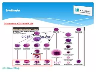
Hemopoiesis and Lymphopoiesis • Pluripotent Stem Cells • Most primitive cells • Mature blood cells and lymphocytes develop from pluripotent cells • The pluripotent stem cells differentiate into either • myeloid stem cells or lymphoid stem cells • The myeloid stem cells produce progenitors • Progenitors lead to the production of mature functional cells RTT 341- Leukemia
Leukemia Development • The development of Leukemia • uncontrolled and accelerated production of progenitors which results in incomplete or defective cell maturation • Acute leukemia- rapid proliferation of primitive, undifferentiated stem cells • Chronic leukemia- differentiated defective cells RTT 341- Leukemia
Leukemia Symptoms • Symptoms result from interference with normal processes • The leukemic cells accumulate in the bone marrow • Hampers the production of normal blood cells • Results in decreased counts What’s this called? RTT 341- Leukemia
Symptoms • Anemia, thrombocytopenia, neutropenia • fatigue • pallor • bleeding • infection RTT 341- Leukemia
ALL-Acute Lymphocytic Leukemia-Epidemiology • 5/100,000 people affected • ALL is the most common pediatric cancer • 80% of children with acute leukemia have ALL • ALL most commonly a childhood disease • Peak incidence between ages 2 and 6 yr old • More common in males, whites compared to blacks, and Jews compared to non-Jews RTT 341- Leukemia
ALL-Acute Lymphocytic Leukemia-Etiology • Unknown causes • Thought to be associated with ionizing radiation-atomic bomb survivors 10-15 % increase incidence • hydrocarbons, benzene, alkylating agents such as cyclophosphamide • Heredity • identical twin has 20% likeliness • Down syndrome- 15-20% increased risk • Naturally occurring retroviruses and the human T-cell lymphotropic virus • -adult ALL- not childhood ALL RTT 341- Leukemia
ALL- Acute Lymphocytic Leukemia Prognosis • Complete remission – more then 75%, duration and therefore “cure” are related to a number of factors: • age; ALL in children < 2 and > 10 poor prognosis. < 1 yr old worst prognosis • Adult ALL worse, > 50 yrs old very poor prognosis • WBC (leukocyte)- initial WBC count 0f < 10,000/mm3 is more favorable than a count of 20,000 to 49,000 and a count > 50,000/mm3 is least favorable RTT 341- Leukemia
ALL- Acute Lymphocytic Leukemia • Clinical presentation • suppressed blood counts - anemia, thrombocytopenia, neutropenia and associated symptoms • flulike malaise with fatigue and pallor from anemia • Thrombocytopenia- bleeding gums, epistaxis , petechiae (tiny red spots on the skin caused by escaped blood), ecchymoses (discolored skin due to blood in tissues), menorrhagia, excessive bleeding after dental procedures RTT 341- Leukemia
ALL- Acute Lymphocytic Leukemia • Clinical presentation: • Neutropenia causes increased risk of infections, respiratory, dental, sinus, perirectal, and UTI • Other common symptoms: • liver, splenic, and testicular enlargement • may mimic rheumatoid arthritis with swollen joints, bone pain, and tenderness causing a child to limp or not walk • Central nervous symptoms-vomiting, headaches, papilloedema (optic disc swelling) neck stiffness, and cranial nerve palsy • ALL- symptoms rarely occur more than 6 weeks before diagnosis RTT 341- Leukemia
ALL-Acute Lymphocytic Leukemia • Detection/Diagnosis • Blood count • thrombocytopenia and anemia occur 2/3 cases at diagnosis • WBC vary • Abnormal increase in WBC poor prognosis • Immunophenotyping • morphological evaluation, special stains, electron microscopy, and surface markers • Establish diagnosis 90% of cases RTT 341- Leukemia
ALL-Acute Lymphocytic Leukemia • Detection/Diagnosis • A bone marrow biopsy is done to make a definitive diagnosis • The amount of leukemic blast cells is determined; <25% is positive for leukemia • Other abnormalities • hyperuricemia, hypomagnesemia, hypocalcemia, and hypercalcemia • 30% of pts have low serum levels of immunoglobulins (proteins that can act as antibodies) RTT 341- Leukemia
ALL-Acute Lymphocytic Leukemia,Detection/Diagnosis • Liver, periosteum, bone may have leukemic infiltrates • Mediastinal mass may be present- high risk pt. • Two most common sites for extramedullary leukemia are • CNS • Testes RTT 341- Leukemia
ALL-Acute Lymphocytic Leukemia • Pathology • ALL is characterized by the uncontrolled proliferation of lymphoblasts • The overproduction of lymphoblasts limits the production of other cells by overcrowding and inhibits cell growth and differentiation • Staging/classification • based on French-American-British (FAB) system. Classified according to cell size, nuclear shape, number of nuclei, prominence of nuclei, and amount of cytoplasm • Levels L1-L3 • 3 being the worse prognosis. Majority of pediatric are L1. RTT 341- Leukemia
ALL-Acute Lymphocytic Leukemia • Staging/classification • 25% of patients with ALL are categorized by T-cell, B-cell, or pre B-cell markers • 70% are classified with null-cell type • this null-cell type reacts to antibody made from an antigen found in ALL cells • antigen is called CALLA, or common leukemia-associated antigen RTT 341- Leukemia
ALL-Acute Lymphocytic Leukemia • Treatment Techniques • Used alone or in combination- • Radiation Therapy (RT)- various techniques • TBI- in prep for BMT • Brain, testes, or CNS (CSI) may be tx with more radiation depending on involvement • TBI- 12Gy, 2 Gy/fx BID 3 days • CNS- helmet (brain) 18 Gy, 2Gy/day, 9 days • CSI- Brain and spine (tx csf) brain- 24Gy, spine 15 Gy • Testes- 4 Gy , 1 tx- often used w/TBI RTT 341- Leukemia
ALL-Acute Lymphocytic Leukemia • Treatment Techniques-cont • Chemo- 3 stages of drugs; induction, consolidation, and maintenance. Protocols change and vary by institutions. • Bone Marrow Transplant-no longer experimental for certain diagnosis and is tx of choice for ALL, AML, and CML RTT 341- Leukemia
ALL-Acute Lymphocytic Leukemia • Side Effects • Acute and Temporary • TBI • GI-nausea, vomiting, diarrhea, anorexia, malaise • Mucosa of mouth, pharynx, bladder and rectum may be affected • Skin reactions, alopecia, interstitial pneumonitis • Decreases blood counts RTT 341- Leukemia
ALL-Acute Lymphocytic Leukemia • Side Effects • Chronic • permanent sterility • cataracts • hepatic fibrosis and radionecrosis of genital tissue, muscle, and kidney • secondary malignancy • lung problems • heart problems • retarded growth RTT 341- Leukemia
AML-Acute Myelogenous Leukemia • Epidemiology • incidence is 5 times greater than ALL • Occurs equally at all ages, slightly more common >50 • 80% of adult leukemia is AML • slightly more common in males • Etiology • Same as for ALL- prior exposure to radiation, benzene, alkylating agents, Fanconi’s anemia, Bloom syndrome (genetic chromosome disorders) RTT 341- Leukemia
AML-Acute Myelogenous Leukemia • Prognosis • prognostic indicators similar to ALL • unfavorable prognosis if: • > 50 yr.s old • Myelodysplastic syndrome- dx of elderly, preleukemia, stem cell disorder • poor performance status • low serum albumin RTT 341- Leukemia
AML-Acute Myelogenous Leukemia • Prognosis • Children w/AML have poorer prognosis than w/ALL • WBC < 20,000, mm is more favorable the 20-49,000 mm, > 50 worst prognosis • Age, tumor burden at time of diagnosis, drug sensitivity of cells are more important prognostic indicators than cell morphology RTT 341- Leukemia
AML-Acute Myelogenous Leukemia • Clinical Presentation • abrupt onset (1-6 month prodromal period) • Similar symptoms to ALL-fatigue, flulike, bleeding, petechiae, purpura (hemorrhage under skin), epistaxis, gingival bleeding, GI bleeding, urinary tract bleeding due to decreased platelets • increased susceptibility to infections due to neutropenia • Enlarged spleen may be felt RTT 341- Leukemia
AML-Acute Myelogenous Leukemia • Detection and Diagnosis • blood counts, abnormal blood counts lead to the detection of AML • Thrombocytopenia, anemia, increased leukocytes • chromosomal abnormalities-30-50% of AML pt • Presence of Auer rods (structures found in myeloblasts, myelocytes, and monoblasts) • bone marrow aspiration biopsy for a definitive diagnosis RTT 341- Leukemia
AML-Acute Myelogenous Leukemia • Detection and Diagnosis • if 30% blast cells are present, acute leukemia is confirmed • a differential diagnosis is made from a staining procedure • Immunophenotyping as in ALL establishes diagnosis in 90% of cases RTT 341- Leukemia
AML-Acute Myelogenous Leukemia • Pathology • proliferation of precursor cells that have lost the ability to differentiate • It involves the hemopoietic stem cells or pluripotent cells • results in the gradual accumulation of undifferentiated cells in marrow or other organs RTT 341- Leukemia
AML-Acute Myelogenous Leukemia • Staging and Classification: • FAB system is used for morphological evaluation • Maturation states are categorized from M0 (undifferentiated) to M7 (megakaryocyte) RTT 341- Leukemia
AML-Acute Myelogenous Leukemia • Treatment techniques • a combination of chemo, radiation therapy, and bone marrow transplant • Side effects-same as ALL RTT 341- Leukemia
CLL-Chronic Lymphocytic Leukemia • CLL most common leukemia • it accounts for 30% of leukemias • CLL is 2 X as common as CML • incidence increases with age, 65 avg, rare in people under 35 • males more common- 2X compared to women • equal blacks/whites RTT 341- Leukemia
CLL-Chronic Lymphocytic Leukemia • Etiology • heredity- 3x increased risk if 1st relative has CLL • Most notable familial clustering of all leukemias • Immunodeficiency syndromes and viruses • no conclusive link with radiation exposure or retroviruses • Prognostic Indicators • stage at time of diagnosis • age • doubling time of peripheral blood lymphocyte count • pattern of bone marrow involvement • T-cell variety poorer prognosis RTT 341- Leukemia
CLL-Chronic Lymphocytic Leukemia • Clinical presentation • incidental findings on blood tests • lymphocyte counts > 10,000/ mm • often asymptomatic • night sweats, fatigue, fever, weight loss • Lymphadenopathy(?) may be present, spleen almost always enlarged • uncomfortable neck masses are common at later stages RTT 341- Leukemia
CLL-Chronic Lymphocytic Leukemia • Detection/Diagnosis • blood tests • Pts always exhibit lymphocytosis • anemia, thrombocytopenia • B-cell origin in 95% of CLL cases • enlarged LN • enlarged spleen • 50% of pt have chromosome abnormalities RTT 341- Leukemia
CLL-Chronic Lymphocytic Leukemia • Pathology • origin may be bone marrow lymphoid tissue • increased number of leukemic cells in bone marrow, blood, lymph nodes, spleen resulting in enlarged spleen, and decreased bone marrow function RTT 341- Leukemia
CLL-Chronic Lymphocytic Leukemia • Staging/Classification • Rai’s staging system (one system) • Three major prognostic indicators are: • Stage 0- low risk • Stages I and II- Intermediate risk • Stages III and IV- high risk • Stages are based on presence of adenopathy, splenomegaly, anemia, and thrombocytopenia. The majority of patients are in the intermediate risk group RTT 341- Leukemia
CLL-Chronic Lymphocytic Leukemia • Staging/Classification • The Binet is another staging system that is based on involvement of cervical nodes, axillary nodes, inguinal nodes, spleen, and liver • CLL- subtypes • Classified as B-cell or T-cell RTT 341- Leukemia
CLL-Chronic Lymphocytic Leukemia • Treatment Techniques • no optimal treatment • early stage pt- no tx benefit • chemo used to treat anemia, thrombocytopenia • radiation used to treat palliatively for localized tumors of lymph tissue • surgery used to remove spleen because of cytopenia, cells accumulate in the spleen RTT 341- Leukemia
CML-Chronic Myelogenous Leukemia • CML accounts for 20-30% of all leukemias • rare in children • uncommon before age 21 • peaks mid 40’s • males slightly more common RTT 341- Leukemia
CML-Chronic Myelogenous Leukemia • Etiology-unknown • linked to radiation, benzene • Philadelphia chromosome is present in 95% of CML patients • Abnormal Chromosome 22- loss of part of long arm • Prognostic Indicators dependent on: • spleen size, platelet count, hematocrit (% of erythrocytes in blood volume), gender, % of blood myeloblasts (immature BM cell) • Can turn into an acute leukemia after 3 yrs-blast crisis • Active phase- 2yr survival RTT 341- Leukemia
CML-Chronic Myelogenous Leukemia • Clinical presentation • three stages • chronic, accelerated, and acute phase (blast crisis) • Early phase- sx are insidious, mild and nonspecific; malaise , fatigue, sweating, intolerance to heat, easy bruising • Splenic enlargement- vague discomfort in LUQ, early satiety, wt loss, peripheral leg edema • Blast crisis 3-4 yrs, all organs are invaded by the leukemic blast cells • symptoms include: fever, bone pain, weight loss RTT 341- Leukemia
CML-Chronic Myelogenous Leukemia • Active disease • Wt. loss > 10% in < 6 months • Fever • Extreme fatigue • Anemia • Thrombocytopenia • Organ involvement (other than LN, spleen, liver, bone marrow) • Progressive or painful enlargement of spleen RTT 341- Leukemia
CML-Chronic Myelogenous Leukemia • Detection Diagnosis • Difficult, insidious, found by accident • Indicators- mild to moderate anemia, leukocytosis • myeloblasts, promyelocytes, and nucleated red blood cells- are present in blood • Absence of LAP score (leukocyte alkaline phosphotase) • BM specimen shows granulocytic and megakaryocytic hyperplasia • Most important–presence of Philadelphia chromosome RTT 341- Leukemia
CML-Chronic Myelogenous Leukemia • Pathology • Abnormal hemopoietic stem cells that give rise to cells that have Philadelphia chromosome • Staging/Classification • Three distinct stages, chronic (stable), accelerated phase, and acute phase (blast crisis) RTT 341- Leukemia
CML-Chronic Myelogenous Leukemia • Treatment Techniques • Radiation • TBI and spleen • Chemo • BMT RTT 341- Leukemia
Role of Radiation Therapist • Radiation departments who do not treat BMT patients or have pediatric oncology services may not see many leukemia patients • Education • Patience- few trips to gain trust • Extra time, preparation (ANESTHESIA) RTT 341- Leukemia
Role of Radiation Therapist • Radiation departments who do not treat BMT patients or have pediatric oncology services may not see many leukemia patients • Education • Patience- few trips to gain trust • Extra time, preparation (ANESTHESIA) RTT 341- Leukemia