Download
1 / 68
680 likes | 760 Views
Explore the intricate network of cells, tissues, and organs comprising the immune system in humans, and learn about the roles of innate and acquired immunity. Discover how lymphocytes like B cells and T cells work to defend against antigens. Dive into the world of adaptive immunity and the vital functions of different immune cells.

E N D
Immune system, lymphoid organs for pharmacists • by Krisztina H.-MinkóSemmelweis UniversityDepartment of Anatomy, Histology and Embryology2018
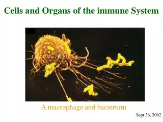
The immune system is a network of cells, tissues, and organs that work together to protect the body from infection. The human body provides an ideal environment for many microbes, such as viruses, bacteria, fungi, and parasites, and the immune system prevents and limits their entry and growth to maintain optimal health. The immune system recognizes and responds to antigens. Antigens are substances (usually proteins) on the surface of cells, viruses, fungi, or bacteria. Nonliving substances such as toxins, chemicals, drugs, and foreign particles can also be antigens. The immune system recognizes and destroys substances that contain antigens. CANCER!
INNATE IMMUNITY Innate, or nonspecific, immunity is the defense system with which you were born. It protects you against all antigens. Innate immunity involves barriers that keep harmful materials from entering your body. These barriers form the first line of defense in the immune response. Examples of innate immunity include: Cough reflex Enzymes in tears and skin oils Mucus, which traps bacteria and small particles Skin Stomach acid Innate immunity also comes in a protein chemical form, called innate humoral immunity. Examples include the body's complement system and substances called interferon and interleukin-1(which causes fever). If an antigen gets past these barriers, it is attacked and destroyed by other parts of the immune system. ACQUIRED IMMUNITY Acquired immunity is immunity that develops with exposure to various antigens. Your immune system builds a defense against that specific antigen.
Lymphocytes B cell: A type of white blood cell and, specifically, a type of lymphocyte. Many B cells mature into what are called plasma cells that produce antibodies (proteins) necessary to fight off infections while other B cells mature into memory B cells. All of the plasma cells descended from a single B cell produce the same antibody which is directed against the antigenthat stimulated it to mature. The same principle holds with memory B cells. Thus, all of the plasma cells and memory cells "remember" the stimulus that led to their formation. The maturation of B cells takes place in birds in an organ called the bursa of Fabricus. B cells in mammals mature largely in the bone marrow. The B cell, or B lymphocyte, is thus an immunologically important cell. It is not thymus-dependent, has a short lifespan, and is responsible for the production of immunoglobulins. It expresses immunoglobulins on its surface. T cell: A type of white blood cell that is of key importance to the immune system and is at the core of adaptive immunity, the system that tailors the body's immune response to specific pathogens. The T cells are like soldiers who search out and destroy the targeted invaders. TCR on the surface. Immature T cells (termed T-stem cells) migrate to the thymus gland in the medistinum, where they mature and differentiate into various types of mature T cells and become active in the immune system in response to a hormone called thymosin and other factors. T-cells that are potentially activated against the body's own tissues are normally killed or changed ("down-regulated") during this maturational process. There are several different types of mature T cells. Not all of their functions are known. T cells can produce substances called cytokines such as the interleukins which further stimulate the immune response (T helper cells, CD4+ cells). Citotoxic T cells (TC cells, or CTLs) destroy virus-infected cells and tumor cells, and are also implicated in transplant rejection. These cells are also known as CD8+T cells since they express the CD8 glycoprotein at their surfaces.
B cell: immunoglobulin production, humoral immune response T cell: TCR receptor, cellular immunity
B cell (soluble antigenes)
MHC-molecules Yoram Reiter Laboratory Department of Biology Technion - Israel Institute of Technology, Haifa 32000, Israel. Projects: TCR-peptide-MHC interaction http://biweb2.bi.technion.ac.il/faculty_websites/reiter/TCR-peptide-MHC%20interaction.htm
T cells: juxtacrin communication T cell receptor T cell http://immbio.aok.pte.hu/hu/oktatas_new/biologus_msc_2011/lectures/biology_msc_immunbiology_9_10_2011.pdf
T lymphocyte functions T helper T citotoxic Connexions; Adaptive Immune Response; Figure 4: Naïve CD4+ T cells engage MHC II molecules on antigen-presenting cells (APCs) and become activated. Clones of the activated helper T cell, in turn, activate B cells and CD8+ T cells, which become cytotoxic T cells. Cytotoxic T cells kill infected cells. http://cnx.org/content/m44821/latest/?collection=col11545/latest
Antigen presenting cells (APC), Dendritic cells MBBS Medicine (Humanity First) / Antigen presenting cells of the skin called dendritic cells http://medicinembbs.blogspot.hu/2011/03/process-of-immunity-images.html
Localisation of lymphocytes in the body http://medical-dictionary.thefreedictionary.com/immune+response
The lymphatic system Primary lymphatic organs are where lymphocytes are formed and mature. They provide an environment for stem cells to divide and mature into B- and T- cells: Both T-cell and B-cells are 'born' in the bone marrow. However, whereas B cells also mature in the bone marrow, T-cells have to migrate to the thymus, which is where they mature in the thymus. Multiplication is not dependent on the presence of antigens. Secondary lymphoid tissues are arranged as a series of filters monitoring the contents of the extracellular fluids, i.e. lymph, tissue fluid and blood. The lymphoid tissue filtering each of these fluids is arranged in different ways. Secondary lymphoid tissues are also where lymphocytes are activated. These include: lymph nodes, tonsils, spleen, Peyer's patches and mucosa associated lymphoid tissue (MALT). Multiplication is dependent on the presence of antigens. http://www.cancerresearchuk.org/ cancer-help/about- cancer/what-is-cancer/ body/the-lymphatic-system
LYMPHOID TISSUE is connective tissue characterized by a rich supply of lymphocytes. Free within the regular connective tissue or is surrounded by capsules, forming the lymphoid organs. Dark blue in HE Rich network of reticular fibers (III. coll) , reticular cells Exception: IN THYMUS : reticulum of unusual epithelial cells Free lymphoid tissue or „encapsulated” Lymphoid infiltration Macrophages, plasma cells
B and T cell precursors Stromal cells Bonemarrow (medullaossium)
THYMUS Bilateral organ Mediastinum T cell maturation Unique structure: epithelium from the 3rd pharyngeal pouches invaded by lymphoblasts
THYMUS HISTOLOGY CT capsule, trabeculae, incomplete lobules, cortex stains more darkly
Cytoreticulum (euchromatic epithelial nuclei) desmosomes Blood-thymus barrier in the cortex Hassall’s corpuscules: keratin, calcification Function? Only efferent lymphatics
Development: from the caudal part of the 3rd pharyngeal pouch
LYMPH NODE/ nodus lymphaticus Bean-shaped, encapsulated structures (2-10 mm) Distributed throught the body along the course of lymphatic vessels In-life filters that are important in the body’s defence against microorganisms and the spread of tumor cells Lymp (tissue fluid) is filtered by at least one node before returning to the circulation
PARTS of the lymph node: CORTEX PARACORTEX MEDULLA Convex surface: entrance for lymph: afferent lymph vessels Concave depression: hilum (efferent lymph vessels, arteries, nerves, veins) Connective tissue capsule, trabeculae Reticular connective tissue http://tpx.sagepub.com/content/34/5/409/F2.expansion.html
folliculus lymphaticus/lymphoid nodules/lymph follicle Unit of the lymphatic tissue primary without germinal center (intrauterin or germ free conditions) secondary follicule(with germinal center). .
folliculus lymphaticus Found in secondary lymphatic organs, or as solitaer lymph follicles (usually under epithelia) In tonsils and mucosa Spherical structures (100 mm. to 1 mm) Contains mainly B cells Site of B lymphocyte activation by antigen bearing macrophage or follicular dendritic cells (FDC) Germinal center (centrum germinativum): site of B lymphocyte proliferation, pale staining Marginal zone LOCATION: Free Lymph nodes Spleen Tonsils NOT in THYMUS!!!!
IN THE CORTEX: reticular cells, macrophages, APCs (dendritic cells, B ly, MHC II on the surface) lymphocytes lymphoid nodules (follicules) interfollicular areas subcapsular sinuses cortical sinuses IN THE PARACORTEX (DEEP CORTEX): NO NODULES (no B cells, but T cells mainly) interdigitating dendritic cells and T cells in venules lymphocytes enter across the vessel wall (HEV) IN THE MEDULLA: medullary cords (B cells, plasma cells, macrophages) medullary sinuses (dilated space between the cords, frequently bridged by reticular cells and fibers)
Lymph flow: aff. lymph vessels, subcapsular sinuses, cortical sinuses, intermediate or trabecular sinuses, medullary sinuses, terminal sinuses. During the passage the lymph is filtered and modified by the immune cells. Finally lymph is collected by the efferent lymphatics.
From arterial end of capillaries fluid is filtered into the extracellular space Fluid gets back to the venous end of capillaries and through lymphatics THE MECHANISM OF LYMPH FORMATION
HISTOLOGY OF LYMPH VESSELS lymphatic capillary valve nucleus of endothelial cell
Largest single accumulation of lymphoid tissue Involved in filtration of blood (defense against blood-borne antigens) Destruction site of erythrocytes Production site of antibodies and activated lymphocytes Any inert particles in blood are actively phagocytosed by spleen macrophages
Spleen structure Capsule (dense CT), trabeculae (subdivide parenchyma, splenic pulp) Hilum (trabeculae: arteries, veins, lymphatics)
Splenic pulp Reticular tissue (reticular cell, lymphocytes, blood cells, macrophages, APCs 2 components: WHITE PULP: lymphoid nodules and periarteriolar lymphoid sheathes „PURPLE STAINING” RED PULP: blood-filled sinusoids and splenic cords (of Bilroth) „REDDISH STAINING”
CIRCULATION IN THE SPLEEN Splenic artery, trabecular arteries, central arterioles (surrounded by PALS, periarteriolar lymphatic sheath), PALS receive large number of lymphocytes (B cells) and may form lymphoid nodules, a. is eccentric but still called central arteriole, smaller branch supply the lymphoid tissue MAGINAL ZONE: surrounding nodules (blood sinuses, lymphoid tissue: lymphocytes, macrophages In the red pulp: penicillar arterioles, splenic cords (network of reticular cells and fibers, lymphocytes, macrophages, plasma cells, erythrocytes, platelets, granulocytes) and venous sinusoids (lined by stave cells, oriented in parallel with the sinusid’s blood flow, sparsely wrapped in reticular fibers set in a transverse direction, like hoops surrounding the staves of a wooden keg)
WHITE PULP: Central arteriole PALS (T cells) Splenic nodules (Malpighian nodules, B cells, a. is eccentric) Marginal zone (loose lymphoid tissue, B cells, marginal sinuses, macrophages).