Download
1 / 95
980 likes | 1.14k Views
Discover the primary and secondary lymphoid organs and their development. Explore lymph nodes, spleen, thymus, and MALT. Learn about the key functions and composition of these vital organs in the immune system.

E N D
Lymphoid Organs Dr. Nabil Khouri MD, MSc, Ph.D
Learning Objectives • Understand the distinction between PRIMARY and SECONDARY lymphoid organs • Be able to describe the general anatomical organization of: • lymph nodes • Spleen • Thymus • Mucosa-associated lymphoid tissue that include: • Diffuse and nodular lymphoid tissue. • regions of extensive lymphoid infiltration such as Peyer’s patches, appendix, and tonsils.
LymphOrgans Free lymphocytes Mucosa-associated lymphoid tissue (MALT). Lymph nodes Tonsils Thymus Spleen
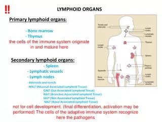
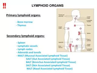
What are Primary lymphatic organs? • Primary lymphatic organs are where lymphocytes are formed and mature. They provide an environment for stem cells to divide and mature into B- and T- cells: • There are two primary lymphatic organs: the Red bone marrow and the Thymus gland. The development of white blood cells (haemopoesis) was covered briefly in the section on blood. • Both T-cell and B-cells are 'born' in the bone marrow. • However, whereas B cells also mature in the bone marrow, T-cells have to migrate to the thymus, which is where they mature in the thymus.
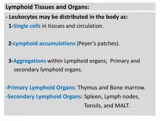
What are Secondary lymphatic organs? • Secondary lymphoid tissues are arranged as a series of filters monitoring the contents of the extracellular fluids, i.e. lymph, tissue fluid and blood. The lymphoid tissue filtering each of these fluids is arranged in different ways. Secondary lymphoid tissues are also where lymphocytes are activated. • These include: lymph nodes, tonsils, spleen, Peyer's patches and mucosa associated lymphoid tissue (MALT).
Development • LYMPH NODE DEVELOPMENT • Except for the upper portion of the cisterna chyli, which persists, the lymph sacs are transformed into groups of lymph nodes during early fetal life, at about month 3. • The surrounding mesenchymal cells invade each sac and break it up into lymphatic channels or sinuses. The mesenchymal cells give rise to the lymph node capsule and the connective tissue framework of the node • The lymphocytes seen in the node before birth come from the thymus gland • The lymph nodule and germinal centers of lymphocyte production do not appear in the nodes until just before or after birth • Lymph nodes also develop along the course of other lymph vessels
Development Other lymph organs • THE SPLEEN develops from an aggregation of mesenchymal cells in the dorsal mesentery of the stomach • THE PALATINE TONSILS form from the second pair of pharyngeal pouches • THE TUBAL (pharyngo-tympanic) TONSILS develop from aggregations of lymph nodules around the openings of the auditory tubes • THE PHARYNGEAL TONSILS (adenoids) develop from an aggregation of lymph nodules in the nasopharyngeal wall • THE LINGUAL TONSILS develop from aggregations of lymph nodules in the root of the tongue
General Lymphoid tissue “Description” • Made up of free cells (Lymphocets, Macrophages and Plasma cells ) • And rich network of reticular fiber • Type III collagen • Dense - Packed with cells • Loose - more reticular fiber fewer cells • Reticular cells produce the fibers
Two Types of lymphoid organs NON ENCAPSULATED (Nodular) MALT (Mucosa Associated Lymphoid Tissue) ENCAPSULATED Direct lymphoid organs Lymph node Spleen Thymus Tonsil • Solitary Nodules • Aggregated nodules (Peyer’s patches) • Lymphoid nodules in vermiform appendix
Mucosa-associated lymphoid tissue (MALT) • MALT is a lymphoid connective tissue located beneath mucous membranes in which the lymphocyte is the predominant cell type. • Occur in the respiratory, gastrointestinal, urinary and reproductive tracts. • The exact extent of these aggregations of lymphocytes mostly B and some T, and helper cells • They have no distinct capsule like that of lymph nodes. • MALT plays a role in regulating mucosal immunity. It may be the site of lymphoma, usually non-Hodgkin lymphoma. • A specific entity is the MALT lymphoma linked to Helicobacter pylori in the stomach.
LYMPHOCYTES IN CONNECTIVE TISSUE: MALT = mucosa-associated lymphoid tissue Diffuse lymphoid tissue within the Lamina propria (LP) of gut and can be found associated with mucosae anywhere in the GIT, Respiratory, and Genitourinary tracts.
MALT: Intraepithelial lymphocytes: Shown here in The GIT – In which T-cells first to see antigens
Intraepithelial lymphocytes Shown here in resp. epithelium.
Nodular Lymphoid Primary Tissue Composition: oval concentrations of lymphocytes contained in meshwork of reticular cells. • Not capsulated • Presented within the lamina propria and mucosa of the organs • Group of B-Lymphocytes arrange in spheres • Called “lymphoid nodules” (Follicles) • When activated by Antigaen-carrying APCs and recognized by B lymphocytes they proliferate in the center called “germinative Center” • This center contain follicular dentritic cells with processes
LN contain follicular dendritic cells Nodule — A small solid collection of tissue, a nodule is palpable (can be felt). It may range in size from greater than 1.0 cm (3/8 inch) to somewhat less than 2 cm (13/16 inch) in diameter. A nodule may be present in the epidermis, dermis or subcutis Primary lymphatic nodule/follicle (LN) Aggregation of lymphocytes in lamina propria or submucosa
Secondary follicles/nodules Composition: • Germinal center - a central region that contains large lymphocytes, mitotic figures, macrophages, and plasma cells. • An outer ring of small lymphocytes. • Arise when B-lymphocytes are presented with appropriate antigen, receive T-cell help, and then begin proliferating as lymphoblasts • Lymphoblasts differentiate into plasma cells or memory cells; aberrant lymphoblasts undergo apoptosis. • Function: morphologic indication of lymphatic tissue response to antigen that represents a cascade of events that includes proliferation of lymphocytes, differentiation of plasma cell, and antibody production.
After the antigen presentation and T-cell help, the activated B-cells set up germinal centers in secondary follicles
The Appendix • Blind sac extending • from the caecum • primary and secondary follicles in lamina propria and submucosa • So, clearly a secondary lymphoid organ… • However, also a site of antigen-INDEPENDENT differentiation • So, also it could be considered as primary lymphoid organ
So, associated with just about any mucosa (GI, respiratory, genitourinary), you may see: • Intraepithelial lymphocytes (T-cells) • Diffuse lymphoid tissue: • B-cells • T-cells • Primary nodules • Secondary nodules • Germinal center with lymphoblasts and mphages
Microfold, or “M” CELLS Are found in the gut-associated lymphoid tissue These cells are modified intestinal epithelial cells that assist in antigen presentation by conveying macromolecules from the intestinal lumen to underlying compartments housing lymphocytes and macrophages.
M cells: TEM M cells are distinguished from other intestinal epithelial cells by their morphological differences. They are characterized by their short or no microvilli. When they present, the microvilli, they are short, irregular, and present on the apical surface or pocket-like invagination on the basolateral surface of these cells.
Distribution of MALT • In the digestive system: • In the wall of the pharynx - tonsils (palatine, lingual, pharyngeal) • In the wall of the small intestine - aggregate lymphoid nodules (Peyer's Patches) or M cells • In the wall of the colon-aggregate lymphoid nodules • In the walls of the appendix • In the reproductive syst. • In the wall of the vagina
Peyer patches are round or oval and are located in the mucous membrane lining of the intestine. They can be seen by the naked eye as elongated thickened areas, and their surface is free of the projections (villi) and depressions (Lieberkühn glands) that characterize the intestinal wall. Usually there are only 30 to 40 patches in each individual. In young adults they may be more numerous, and as a person ages they tend to become less prominent. Their full function is not known, but they do play a role in immunologic response and contain B and T cells similar to those found in peripherallymph nodes.
Peyer’s patches • Peyer’s patches are roughly egg-shaped lymphatic tissue nodules that are similar to lymph nodes in structure, except that they are not surrounded by a connective tissue capsule. They belong to a class of Non-Encapsulated lymphatic tissue known as lymphatic nodules, which include the tonsils and lymphatic tissue of the appendix
Peyer’s patches Peyer’s patches are found throughout the ileum region of the small intestine known as aggregated lymphoid nodues, They form an important part of the immune system by monitoring intestinal bacteria populations and preventing the growth of pathogenic bacteria in the intestines.
Tonsils This lymphatic tissue belong to Mucosa-associated Lymphoid Tissue (MALT) group. They are considered organs because they are partially encapsulated Tonsils are covered by an epithelium depending on their location They include: 1. Palatine tonsils 2. Pharyngeal tonsils 3. Lingual tonsils 4. Tubal tonsils
lymphoid organs Tonsils - A paired Lymphoid structure located in the Oropahrynx trap and destroy bacteria
Palatine Tonsils • The non-capsulated surface is covered contains Dense lymphoid tissue (follicles) that forms a band of lymphatic nodules that lie below the stratified squamous epithelium lining the oral cavity in this region. • Subdivided into lobes by 10-20 crypts
Palatine Tonsils Palatine tonsils • Overlying epithelium forms invaginations called multiple crypts that penetrate into the band of nodules. • These crypts act as collecting places for cellular debris and bacteria as well as some living lymphocytes that have migrated into the crypts. • The band of lymph nodules is separated from underlying tissues by a partial capsule of dense connective tissue.
Pharyngeal Tonsil • Located in the Naso-pharynx • Covered by ciliated Pseudostratified epithelium • In Some areas of the covering epithelium may be stratified squamous. • Form a thin sheet of lymphoid nodules and diffuse lymphocytes • Diffuse lymphoid tissue and nodules, but no crypts. • Thin partial capsule of dense connective tissue separates the lymphoid tissue from underlying tissue. • Chronic inflammation = Adenoid
Pharyngeal Tonsil Tonsil that has nodules and covered by psedostratified epithelium with No Cripts ⇒ Pharyngeal tonsil
Lingual Tonsils They are multiple small collections of lymphoid tissue located at the base of the tongue Lingual Tonsil are Covered by Non keratinized stratified squamousepithelium One crypt for each tonsil Or without deep crypts ⇒ Lingual tonsil.
Along the course of lymphatic vessels there are numerous small Bean shaped structures called LYMPH NODES • Usually present in groups (will be presented for you in a separate session) • Lymph from any part of the body passes through one or more lymph nodes before entering the blood stream • Lymph nodes act as filter removing bacteria and other particulate matter from lymph • Provides necessary microenvironment for antigen-dependent differentiation • Lymphocytes are added to lymph in these nodes
Anatomy of lymph nodes: • Entire node is Bean shaped • The concavity constituting a Hilum • Usually a single lymph vessel leaves the node through its hilum. • Several lymph vessels enter the node on its convex aspect • Each lymph node consists of, • Connective tissue framework • Lymphocytes
Lymph Node Structure The Capsule & Subcapsular sinus Send Trabeculae & trabecular sinuses Sinuses contain lymph, macrophages, and reticular cells • The Cortex: • An Outer “superficial” Part (B-cells) contains: • -primary follicles/nodules • -secondary follicles/nodules • An Inner “deep” Part (T-cells, dendritic cells) • - The Medulla: • medullary cords (B-cells, plasma cells) • medullary sinuses (lymph, more macrophages, plasma cells, and reticular cells)
The Medulla • The medulla of a lymph node is composed of • medullary cords interspersed between medullary sinuses. • The medullary cords are composed of dense lymphoid tissue contain primary B lymphocytes their precursors plasma cells, macrophages and T helper cells. • The most prominent cell in the cord is the precursor to plasma cells or immunoblasts that came from the germinal centers of the lymphoid follicles in the cortex of the node. • The medullary sinuses are composed primarily of reticular fibers (RF) providing the support framework, reticular cells (fibroblast-like cells that secret the reticulin). • Contain lymph , lymphocytes and macrophages
The cortex • Is composed of the cortical sinuses surrounded by dense accumulations of lymphocytes. • In the more superficial cortex the lymphocytes are arranged into spherical follicles, lymphoid follicles where B lymphocytes are activated and undergo proliferation. • GERMINAL CENTER (GC) contains pale-staining cells. • The open, pale-staining nature of the nuclei of these cells indicate that they are T and B lymphocytes undergoing active proliferation. • Other cells include: • Reticular cells = follicular dendritic cells that present antigen to the B Lymphocytes • Macrophages that engulfed dead B cells that have died by apotosis
Para-cortical zone, Sub-capsullar and Radial sinuses • Subcapsullar regions of the cortex is made of loose lymphoid tissue with reticular cells and fibers • Radial sinuses of the cortex is placed between the nodules and Contain primarily T lymphocytes that do not form into follicles. • T lymphocytes enter the lymph node parenchyma reside in the Paracortical zone. • If activated, the T lymphocytes undergo active proliferation to produce expanded clones of activated T lymphocytes. Capsule Sub-capsular sinuses Radial sinuses
From the sub-capsular sinus, lymph percolates through trabecular sinuses, and finally into MEDULLARY SINUSES
High magnification view of a sinus (subcapsular sinus shown here) M=macrophage, Ly=lymphocytes, RF/RC=reticular fiber (and associated reticular cell)
Micrographs of lymph node of a cat showing medullary sinuses and cords.
silver impregnation to visualize Reticular Fibers Special stain: • RF Form a delicate supporting framework for highly cellular tissues • found in lymph nodes, liver, bone marrow, spleen, smooth muscle). • Composed mainly of Type III collagen. • Thinner than type I collagen