Download

1 / 15
200 likes | 509 Views
Histological Structure of Lymphoid Organs. DR RANIA GABR. Objectives. Understand the location of lymphatic organs. Discuss the microscopic features of Lymph Node. Discuss the microscopic features of Spleen. Discuss the microscopic features of Thymus.

E N D
Histological Structure of Lymphoid Organs DR RANIA GABR
Objectives • Understand the location of lymphatic organs. • Discuss the microscopic features of Lymph Node. • Discuss the microscopic features of Spleen. • Discuss the microscopic features of Thymus. • Discuss the microscopic features of Tonsils
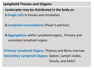
Lymphoid Tissue • Lymphoid tissue is connective tissue chch by rich supply of lymphocytes. • It is found either 1- Free in regular CT 2-Surrounded by capsules, forming the “lymphoid organs” • Very little cytoplasm so stain dark blue with H&E. • Rich network of reticular fibrils produced by fibroblasts.
Lymphoid System Basics • Two main tissue architecture types: • Diffuse: uniform appearance • Follicular: consists of lymphoid follicles • Two types of lymphoid tissues: • Encapsulated: connective tissue capsule • spleen, thymus, lymph nodes • Unencapsulated (or partly encapsulated) • Tonsils, Peyer’s patches, lymphoid nodules in GI tract, respiratory tract, urinary & reproductive tracts
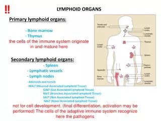
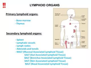
2 Types of Lymphoid Organs • Central (primary) lymphoid organ: where lymphoid cells undergo maturation • T cells in thymus • B cells in bone marrow • Peripheral (secondary) lymphoid organ: where functional lymphocytes go including: 1- lymph nodes 2- spleen, 3- Peyer’s patches, 3- lymphoid nodules of GI and other tracts
Lymphoid Follicles • Nodules of densely packed lymphocytes located in all peripheral lymphoid tissues. Most lymphocytes are B cells. • Two distinct areas: 1- Mantle– darker stained, mainly small, resting lymphocytes 2- Germinal center – (defines “secondary” or “reactive” lymphoid follicles): lighter stained, larger, activated B cells
Lymph follicle: • Mantle = cap (dark) • Germinal center (light)
Lymph Nodes • Present throughout the body, along lymph vessels • Numerous in axilla, groin, cervical area and thoracic/abdominal mesenteries • Filter lymph before it returns to vasculature • Hilum: concave side, arteries, nerves enter; veins and efferent lymph vessels leave the organ • Afferent lymph vessels enter convex surface
Covered by a capsule which extends to form Trabeculae. • Divided into outer cortex and inner medulla. • OUTER CORTEX contains: Lymphatic nodules with germinal center • INNER MEDULA contains: Medullary Cords and Medullary Sinus
Medullary cords • Are branched, cordlike extensions of lymphoid tissue arising from the paracortex. They contain primarily B- lymphocytes and often plasma cells and macrophages. • Medullary cords are separated by dilated spaces , frequently bridged by reticular cells and fibers , called Medullary sinuses • They contain lymph , lymphocytes, macrophages, sometimes granulocytes if the lymph node is draining an infected organ
Lymph nodeCT --- Connective tissueC --- CortexM --- MedullaP --- ParacortexLN --- Lymph NodeT ---TrabeculaeMS --- Medullary SinusMC --- Medullary Cords