Download
1 / 54
550 likes | 823 Views
Tissues and Organs Comprising the Immune Response System. Where components of the immune response originate, differentiate, meet, interact, and function. Updated: October 30, 2013. Folder Title: Tiss&OrgNoTP. APC and T-Cells. Marcophage and T-Cell Kissing.

E N D
Tissues and OrgansComprising the ImmuneResponse System Where components of the immune response originate, differentiate, meet, interact, and function Updated: October 30, 2013 Folder Title: Tiss&OrgNoTP
Helper T-cell B-Cell Interaction CD4 co-receptor
B-Cell Activation See figure 2-7, Kuby, 4th Ed., p. 36
B-Cell: Small, Blast, and Plasma See Figure 2-7, Kuby 4th Ed. p. 36
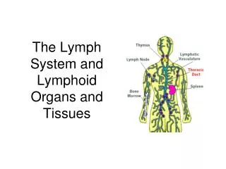
Where do these cells originate? Where do they go to interact with each other? Where do they carry out their functions?
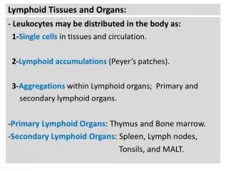
Related to Figure 2-13, Kuby, 4th Edition, p. 47 Primary and Secondary Lymphoid
Animations Produced for Kuby Immunology http://bcs.whfreeman.com/immunology6e/ Chapter 2 Animation. Cells and Organs
Please put away all notes and any devices except for your Turning Point NXT Transmitter. No papers or computers on your desk please. No communication between or among students.
Thymus Cartoon Diagrammatic Cross Section of Thymus. Figure 2-14, Kuby, 4th Edition, p. 48 ThymusCross
See Figure 2-22, Kuby 4th Ed. p. 56 MALT and M-Cells
MALT and IgA See Figure 2-22 Kuby, 4th Ed. p. 56
CD Antigen Table CDMarks
Animations Produced for Kuby Immunology http://bcs.whfreeman.com/immunology6e/ Chapter 11 Animation. Signal Transduction
CD Antigen List: Pages A1 to A26: 339 Entries, 2007 Edition of Kuby How to Understand CD Antigens: 5 Persons in a Room Person 1: 5 ft 5”, 160 lbs bespectacled old geezer Person 2: 5 ft 11”, 280 lbs, 22-year old guy, all muscle Person 3: 5 ft 5”, 135 lbs, 20-year-old woman Person 4: 7 ft 1” , 200 lbs, 22 year-old guy Person 5: 5 ft 8”, 150 lbs, 22 year-old guy
Person 1: 5 ft 5”, 160 lbs bespectacled old geezer Person 2: 5 ft 11”, 280 lbs, 22-year old guy, all muscle Person 3: 5 ft 5”, 135 lbs, 20-year-old woman Person 4: 7 ft 1” , 200 lbs, 22 year-old guy Person 5: 5 ft 8”, 150 lbs, 22 year-old guy This is a fill-in-the-blank question. Please enter your responses as a series of numbers from 1 to 5. Which Person:A. Is a varsity basketball player?B. Plays on the women’s field-hockey team?C. Is a boring old professor?D. Is a varsity football player for SU?E. Is a cross-country runner?__ __ __ __ __
Secrets of Signaling 1. How Do You Knock on a Door? 2. Why does the phone “ring” more than once? 3. Why does the police officer blow his whistle only once? 4. Why does your computer say “Are You Sure You Want to Delete File XYZ? 5. When Ashley Nieves asked the class to raise their right hand, why did only eleven persons out of 95 respond? 6. Why did a different set of students respond to the same message when InSu Jo made the request?
T-Cell ALL Markers (“ALL” = Acute Lymphocytic Leukemia) Kuby, 5th Edition p. 157
Pre-B-Cell ALL Markers (“ALL” = Acute Lymphocytic Leukemia) Kuby, 5th Edition p. 157
B-Cell CLL Markers (“CLL” = Chronic Lymphocytic Leukemia) Kuby, 5th Edition p. 157
Membrane of CirculatingLeucocytes General Structures of Cell Adhesion Molecules (CAMs) Kuby, 3rd Edition Figure 15-2a CAMStruct Endothelial Cells Lining Capillaries
Actin Cytoskeleton Linkage to Collagen Extra-cellular Matrix Figure 19-14 ECB 1998, p. 603 Collagen Fibronectin Integrin Plasma Membrane Actin Microfilament MFtoECM
Animations Produced for Kuby Immunology http://bcs.whfreeman.com/immunology6e/ Chapter 13 Animation. Leukocyte Extravasation
On a scale from –2 to +2(Use #1 on Key-pad) -2 = I’m lost; (Use #2 on Key-pad) -1 = I’m having a hard time, but I follow some of it.(Use #3 on Key-pad) 0 = I’m doing OK. I uderstand most of it. I can figure the rest out later.(Use #4 on Key-pad) +1 = I’m following OK. No problem(Use #5 on Key-Pad) +2 = This is pretty straight-forward, Please move on.