Water, Electrolyte &
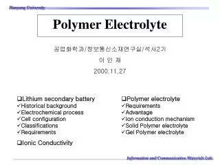
Prof. Mehdi Hasan Mumtaz. Water, Electrolyte &. Acid-Base Balance. BALANCE. Water Balance Electrolyte Balance. Acid Base Balance. Nutritional Balance. IVS. ISS. ICS. 5L. 14L. 23L. TOTAL BODY WATER 42L. FLUID THERAPY. INTRACELLULAR INTERSTITIAL VASCULAR. CAPILLARY. EG.

Water, Electrolyte &
E N D
Presentation Transcript
Prof. Mehdi Hasan Mumtaz Water, Electrolyte & Acid-Base Balance
BALANCE • Water Balance • Electrolyte Balance. • Acid Base Balance. • Nutritional Balance.
IVS ISS ICS 5L 14L 23L TOTAL BODY WATER 42L
FLUID THERAPY INTRACELLULAR INTERSTITIAL VASCULAR CAPILLARY EG CELL OSMOLALITY Na+ COP
ACID-BASE BALANCE • Terminology. • Physiologic Compensation By Body. • Pathophysiologic Disturbances. • Practical Approach To Assessment. • Biochemical Reports & Case Histories.
DEFINITION OF TERMINOLOGY • ACID - STANDARD BICARBONATE. • BASE - BUFFER BASE & BASE DEFICIT. • ALKALI • BUFFERING & BUFFER. • PH. 24 x PCO2 (mmHg) H+(nmol/L)=- ----------------------------- HCO3 (meq/L) (40nmol/L)
PRODUCT OF METABOLISM • H++ - Anaerobic Metabolism. • CO2 - Aerobic Metabolism.
PHYSIOLOGIC COMPENSATION • HYDROGEN IONS. • Incoporation in water.H++HCO3H2C3O CO2 + H2O. • Loss from body. • Kidney – regeneration of HCO3. • Intestine. • CO2. • Chemoreceptors in hypothalamus. • HCO3. • HCO3 generation by erythrocytes. • HCO3 re-absorption in renal tubules. • HCO3 generation in renal tubules.
BICARBONATE GENERATION BY ERYTHROCYTES HHB Hb Cl— HCO-3 CO2 Cl— -HCO3 +H+ CO2+H2O
HCO3- Na+ Na+ HCO3- HCO3- HCO3- H+ CELL H2CO3 H2CO3 CD CO2 H2O CO2 + H2O BICARBONATE REABSORPTION BY KIDNEY RENAL T. LUMEN STIMULATED BY HCO3- M. ACIDOSIS
BICARBONATE GENERATION IN KIDNEY B- HCO3- Na+ STIMULATED PCO2 (BY RESP ACIDOSIS) & -HCO3 (M. ACIDOSIS) Na+ B- HCO3- H+ CELL H2O HB CO3 H2O
PATHOPHYSIOLOGIC DISTURBANCES Lungs Disturbances of CO2= R. Centre Disturbance of H++HCO3 = Metabolic
Henderson - HosselbalchEQUATION Proton Acceptor (Base) PH=PK+Log = -------------------------------- Proton Donor (Acid) -HCO3 (Metabolic) PH=PK+Log = ---------------------------------- H2CO3 or PCO2 x 0.03 (Respiratory)
ACID-BASE DISTURBANCE -HCO3 PCO2 x 0.03 MEATBOLIC RESPIRATORY ACIDOSIS ALKALOSIS ACIDOSIS ALKALOSIS HCO3 ---------------- PCO2x0.03 HCO3 ---------------- PCO2x0.03 HCO3 ---------------- PCO2x0.03 HCO3 ---------------- PCO2x0.03 RATIO
Metabolic acidosis = Respiratory acidosis = Metabolic alkalosis = Respiratory alkalosis = Defect HCO3 ---------- PCO2 HCO3 ---------- PCO2 HCO3 ---------- PCO2 HCO3 ---------- PCO2 Correction HCO3 ---------- PCO2 HCO3 ---------- PCO2 HCO3 ---------- PCO2 HCO3 ---------- PCO2
CAUSES OF M. ACIDOSIS Hyperkalamic M. Acidosis • Glomeralar failure. • Keto-acidosis. • Lactic acidosis. • Intestinal loss. • R. Tubular failure. • Actazolamide therapy. • R. Tubular acidosis. • Ureteric transplantation. • NH4Cl el therapy. Variable Hyppkalamic Acidosis Hyperchloraemic Acidosis
SCREENING TESTS METABOLIC ACIDOSIS • BLOOD GLUCOSE. • URINE/ BLOOD KETONES. • SERUM CHLORIDE. • SERUM POTASSIUM
RESPIRATORY ACIDOSIS • Acute Respiratory Failure. • Erythrocyte • Chronic Respiratory Failure. • Renal Generation.
METABOLIC ALKALOSIS • Administration of HCO3. • K+ depletion – Generation by kidney. • Pyloric Stenosis.
RESPIRATORY ALKALOSIS • Hysterical Over-breathing. • ICP. • Brain Stem Injury. • Hypoxia. • Pulmonary Oedema. • Lobar Pneumonia. • Pulmonary Collapse. • Excessive Artificial Ventilation.
BALANCE OF ACID-BASE NORMAL VALUES • PCO2 • 30-50mmHg or 4-6.6kPa. • >50mmHg respiratoryor 6.6kPa acidosis • <30mmHg respiratoryor 4kPa alkalosis • PH • 7.30 – 7.50 • >7.50 alkalaemia. • <7.30 acidosis
BALANCE OF ACID-BASE RELATIONSHIP • PCO2 and PH. • PCO2 &ventilation. • PO2 andnormal range. • PO2 and FIO2. • PCO2,and temperature.
TERMINOLOGY • ACIDAEMIA - PH<7.30 • ALKAEMIA - PH>7.50. • ACIDOSIS - Base Deficit Present. • ALKALOSIS - Base Excess Present.
HOW TO ASSESS BLOOD GASES? • STEP-1 Assessment of Acid-Base Balance. • STEP-2 Assessment of Hypoxaemic State. • STEP-3 Assessment of Tissue Oxygenation State.
STEP-1Assessment of Acid-Base BalanceCLASSIFICATION ALKALOSIS ACIDOSIS METABOLIC RESPIRATORY METABOLIC RESPIRATORY CHRONIC ACUTE CHRONIC ACUTE CHRONIC ACUTE CHRONIC ACUTE
STEP-1Assessment of Acid-Base Balance • Acute - Uncompensated. • Chronic - Compensated. -Fully. - Partially. COMPENSATED PH 7.30-7.50 DIAGNOSIS.
DIAGNOSIS SEQUENCE. • PH. • PCO2. • HCO3. PH Normal 7.4 Compensated 7.3-7.5 PCO3 Normal 40mmHg (5.3kPa) Compensated 30-50mmHg (4-6.6 kPa)
DIAGNOSIS • IF PH LOW – acidosis. • Look at PCO2. • If PCO3 high - respiratory acidosis • If PH low - acidosis • Look at PCO2 • If it is normal or low. • Look at HCO3. It is low – metabolic acidosis. • IF PH HIGH - alkalosis • Look at PCO2. • If it is low - respiratory alkalosis • If PH high - PCO2 normal or high. • Look at HCO3. High - metabolic alkalosis. NOW LOOK FOR COMPENSATION
A C I D O S I S A L K A L O S I S Primary change Primary change
STEP-2Hypoxaemic State • Below 60 years of age: • Normal PO2 = 97mmHg. • Acceptable range = >80mHg. • Mild hypoxiaemia = <80mmHg. • Moderate hypoxiaemia = <60mmHg. • Severe hypoxiaemia = <40mmHg.
STEP-2Hypoxaemic State • Above 60 years of age: • Subtract 1mmHg from minimal 80mmHg for every year over 60; this means acceptable range: • 60 years = >80 mmHg. • 70 years = >70 mmHg. • 80 years = >70 mmHg. • 90 years = >50 mmHg. • New Born: • Acceptable = 40-70 mmHg.
STEP-2Hypoxaemic State • Oxygen Therapy FIO2 x 5 = Expected PO2. Uncorrected Hypoxaemia = PO2<Room Air Acceptable Limit. Corrected Hypoxaemia = PO2 > Room Air Acceptable Limit. <100mmHg. Excessively Corrected Hypoxaemia = PO2>100mmHg < minimal predicted.
STEP-3Assessment of Tissue Oxygenation • Cardiac Status. • Peripheral Perfusion Status. • Blood Oxygen Transport Mechanism. Depends on: • Vital Signs • Physical Examination.
STEP-3Assessment of Tissue Oxygenation • BP. • Pulse Pressure. • Heart Rate • ECG. • Skin Color & Condition. • Capillary Fill. • Senosrium. • Electrolyte Balance. • Urine Out Put. • If Above 1,2 Good Only 3 Interfering. • Arterial Oxygen Tension Po2. • Blood Oxygen Content. • Hb Oxygen Affinity.
SUMMARY • ASSESS ACID/BASE STATUS. • ASSESS HYPOXAEMIC STATE • ASSESS TISSUE OXYGENATION. • TRY TO FIND OUT THE CAUSE. • SEE FOR THE NEED OF HCO3.
SUMMARY Acidosis Metabolic 6. If Cl- K+ Think of actazolamide therapy and R. Tubul Acidosis. 7. If Cl-N K+ Proximal Tubul Failure. OTHERWISE THINK ABOUT GIT INVOLVEMENT Look at 1. Blood urea If and K+ G.F. 2. Blood Glucose ket If and K+ ketoacidosis. 3. PO2 If K+ Lactic acidosis 4. Serum HCO3. If only H/o Therapy 5. If K+ think of NH4Cl therapy + G. Transplantation
SUMMARY Respiratory Alkalosis Lung Functions will Help METABOLIC Look at K+ & Cl- K+ Cl- H/o vomiting Pyloric stenosis If K+ find cause. H/o bicarb therap. RESPIRATORY - H/o H. Injury - L. Infection - IPPV
BASE EXCESS/ DEFICIT “mEq of HCO3 that is excess/ deficit per litre of E. C. Water” PREDICTED RESPIRATORY PH? PCO2 -- PH RELATIONSHIP PCO2 20mmHg = 0.1PH. PCO2 10mmHg = 0.1PH
BASE EXCESS/ DEFICIT • Calculate difference between measured PCO2 and 40mmHg. Move decimal 2 places to left. • If PCO2 > 40 subtract ½ difference from 7.4. • If PCO2 < 40 add the difference to 7.40. • PH 7.21 PCO2 90 • 90-40 = 50 = 0.50 = 0.50x ½ = 0.25 • 7.40-0.25 =7.15 • PH 7.47 PCO2 18 • 40-18 = 22= 0.22 • 7.40 + 0.22 =7.62 Predicted Resp PH.
DETERMINATION OF METABOLIC COMPONENT 10mEq/L variance from buffer base PH change of c-15 units. Move decimal 2 places to right i.e. 15 ratio 15:, 2:3=2/3 Measured PH - Predicted PH (resp) - metabolic PH change.
DETERMINATION OF METABOLIC COMPONENT • Determine PCO2 variance. I.e. PCO2 -40mmHg PCO2. • Move decimal 2 point to left. • Determine Predicted Resp. PH. • Measured PH – Predicted PH difference move decimal 2 places to rt. X 2/3=base excess/deficit. Base Excess = measured PH> predicted PH. Base Deficit = measured PH> predicted PH.
DOES TRADITIONAL BLOOD GAS ANALYSIS SERVES THE PURPOSE? PH PCO2 PO2 HCO3
WHAT INFORMATION DOES IT GIVE? OXYGEN UPTAK CO2 PRODUCTION ACIDITY/ ALKALINITY
WHAT INFORMATION IS REQUIRED FOR THERAPY? • UPTAKE - O2 uptake in lungs. • TRANSPORT - from lungs to capillaries. • RELEASE - from capillaries to tissues. HOW TO WE GET? DEEP PICTURE OF BLOOD GASES
O2 UPTAKE MOUTH TO ALVEOLI “Grahams’ Law” of diffusion
O2 UPTAKE Alveoli to Hb “Henrys’ Law” of diffusion
COMBINE BOTH LAWS • Mouth to Alveoli Grahams’ Law of diffusion • Alveoli to Hb Henrys’ Law of diffusion.
TRANSPORT TO CAPILLARIES DO2 “ 520 - 720ml/min/m2”