Download
1 / 67
690 likes | 1.12k Views
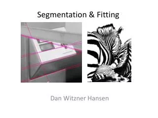
MEDICAL IMAGING INFORMATICS: Lecture # 6 Segmentation. Norbert Schuff Professor of Radiology VA Medical Center and UCSF Norbert.schuff@ucsf.edu. Overview. Definitions Role of Segmentation Segmentation methods Intensity based Shape based Texture based Summary & Conclusion Literature.

E N D
MEDICAL IMAGING INFORMATICS:Lecture # 6Segmentation Norbert Schuff Professor of Radiology VA Medical Center and UCSF Norbert.schuff@ucsf.edu Department of Radiology & Biomedical Imaging
Overview • Definitions • Role of Segmentation • Segmentation methods • Intensity based • Shape based • Texture based • Summary & Conclusion • Literature Department of Radiology & Biomedical Imaging
The Concept Of Segmentation Identify classes (features) that characterize this image! Intensity: Bright - dark Shape: Squares , spheres, triangles Texture: homogeneous – speckled Connectivity: Isolated - connected Topology: Closed - open Department of Radiology & Biomedical Imaging
More On The Concept Of Segmentation Can you still identify multiple classes in each image? Department of Radiology & Biomedical Imaging
Segmentation Of Scenes Segment this scene! Hint: Use color composition and spatial features By J. Chen and T. Pappas; 2006, SPIE; DOI: 10.1117/2.1200602.0016 Department of Radiology & Biomedical Imaging
Gray matter segmentation Segmentation of abdominal CT scan By intensity Stephen Cameron. Oxford U, Computing Laboratory By texture image at: www.ablesw.com/3d-doctor/3dseg.htm Examples: Intensity and Texture Department of Radiology & Biomedical Imaging
Definitions • Segmentation is the partitioning of an image into regions that are homogeneous with respect to some characteristics. In medical context: • Segmentation is the delineation of anatomical structures and other regions of interest, i.e. lesions, tumors. Department of Radiology & Biomedical Imaging
Formal Definition If the domain of an image is , then the segmentation problem is to determine sets (classes) Zk, whose union represent the entire domain Sets are connected: Department of Radiology & Biomedical Imaging
More Definitions • When the constraint of connected regions is removed, then determining the sets Zk is termed pixelclassification. • Determining the total number of sets K can be a challenging problem. • In medical imaging, the number of sets is often based on a-priori knowledge of anatomy, e.g. K=3 (gray, white, CSF) for brain imaging. Department of Radiology & Biomedical Imaging
Labeling • Labeling is the process of assigning a meaningful designation to each region or pixel. • This process is often performed separately from segmentation. • Generally, computer-automated labeling is desirable • Labeling and sets Zk may not necessarily share a one-to-one correspondence Department of Radiology & Biomedical Imaging
Dimensionality • Dimensionality refers to whether the segmentation operates in a 2D or 3D domain. • Generally, 2D methods are applied to 2D images and 3D methods to 3D images. • In some instances, 2D methods can be applied sequentially to 3D images. Department of Radiology & Biomedical Imaging
Characteristic and Membership Functions • A characteristic function is an indicator whether a pixel at location j belongs to a particular class Zk. • This can be generalized to a membership function, which does not have to be binary valued. • The characteristic function describes a “deterministic” segmentation process whereas the membership function describes a “probabilistic” one. Department of Radiology & Biomedical Imaging
Segmentation Has An Important Role Computational diagnostic Surgical planning SEGM Database storage/retrieval Image registration Atlases informatics Image transmission Quantification Partial volume correction Super resolution Department of Radiology & Biomedical Imaging
Segmentation Methods Department of Radiology & Biomedical Imaging
Histogram (fictitious) Threshold (TA) (TB ± ) Threshold Method Angiogram showing a right MCA aneurysm Dr. Chris Ekong; www.medi-fax.com/atlas/brainaneurysms/case15.htm Department of Radiology & Biomedical Imaging
Threshold Method Original Threshold min/max Threshold standard deviation Department of Radiology & Biomedical Imaging
Threshold Method Applied To Brain MRI White matter segmentation • Major failures: • Anatomically non-specific • Insensitive to global signal inhomogeneity Department of Radiology & Biomedical Imaging
Threshold: Principle Limitations • Works only for segmentation based on intensities • Robust only for images with global uniformity and high contrast to noise • Local variability causes distortions • Intrinsic assumption is made that the probability of features is uniformly distributed Department of Radiology & Biomedical Imaging
Seed point Region Growing - Edge Detection • Region growing groups pixels or subregions into larger regions. • A simple procedure is pixel aggregation, • It starts with a “seed” point and progresses to neighboring pixels that have similar properties. • Region growing is better than edge detection in noisy images. Guided e.g. by energy potentials: Similarity: Edges: Department of Radiology & Biomedical Imaging
CT of different types of bone tissue (femur area) Gray scale intensity (a) WS over-segmentation (b) WS conditioned by regional density mean values (c) WS conditioned by hierarchical ordering of regional density mean values M. Straka, et al. Proceedings of MIT 2003 Region Growing – Watershed Technique Department of Radiology & Biomedical Imaging
Region Growing: Principle Limitations • Segmentation results dependent on seed selection • Local variability dominates the growth process • Global features are ignored • Generalization needed: • Unsupervised segmentation (i.e.insensitive to selection of seeds) • Exploitation of both local and global variability Department of Radiology & Biomedical Imaging
Clustering • Generalization using clustering • Two commonly used clustering algorithms • K-mean • Fuzzy C-mean Department of Radiology & Biomedical Imaging
Definitions: Clustering • Clustering is a process for classifying patterns in such a way that the samples within a class Zk are more similar to one another than samples belonging to the other classes Zm, m ≠k; m = 1…K. • The k-means algorithm attempts to cluster n patterns based on attributes (e.g. intensity) into k classes k < n. • The objective is to minimize total intra-cluster variance in the least-square sense: • for k clusters Zk, k= 1, 2, ..., K. µi is the mean point (centroid) of all pattern values j ∈ Zk. Department of Radiology & Biomedical Imaging
Fuzzy Clustering • The fuzzy C-means algorithmis a generalization of K-means. • Rather than assigning a pattern to only one class, the fuzzy C-meansassigns the pattern a number m, 0 <= m <= 1, described as membership function. Department of Radiology & Biomedical Imaging
K- means Three classes Department of Radiology & Biomedical Imaging
K - means Four classes Department of Radiology & Biomedical Imaging
K - means Original Department of Radiology & Biomedical Imaging
K - means 2 clusters Department of Radiology & Biomedical Imaging
K - means Three classes Department of Radiology & Biomedical Imaging
K – means: TRAPPED! Five classes Department of Radiology & Biomedical Imaging
Fuzzy C - means Four classes Department of Radiology & Biomedical Imaging
Fuzzy C- Means Segmentation I Two classes Original Class I Class 2 Department of Radiology & Biomedical Imaging
Class 2 Class I Class 3 Class 4 Fuzzy Segmentation II Four classes Original Department of Radiology & Biomedical Imaging
Brain Segmentation With Fuzzy C-Means 4T MRI, bias field inhomogeneity contributes to the problem of poor segmentation Department of Radiology & Biomedical Imaging
Clustering: Principle Limitations • Convergence to the optimal configuration is not guaranteed. • Outcome depends on the number of clusters chosen. • No easy control over balancing global and local variability • Intrinsic assumption of a uniform feature probability is still being made • Generalization needed: • Relax requirement to predetermine number of classes • Balance influence of global and local variability • Possibility to including a-priori information, such as non-uniform distribution of features. Department of Radiology & Biomedical Imaging
Segmentation As Probabilistic Problem • Treat both intensities Y and classes Zas random distributions • The segmentation problems is finding the classes that maximize the likelihood to represent the image • Segmentation in Bayesian formulation becomes : • where • Y is the observed image (values y1 ….yn) • Z is the segmented image (classes z1 ….zK) • p(Z) is the prior probabillity • p(Y|Z) the observation probability • and p (Y) is the observation and hence stable Department of Radiology & Biomedical Imaging
Treat As Energy Minimization Problem • Since p(Y) is stable, it follows: • The goal is to find the most probably distribution of p(Z|Y) given the data • Since the log probabilities are all additive, they are equivalent to distribution of energy • segmentation becomes an energy minimization problem. • This means in particular that no probabilistic point of view is finally required. Department of Radiology & Biomedical Imaging
Probability In Spatial Context • Use the concept of Markov Random Fields (MRF) for segmentation • Definition: • Classes Z are a MRF on Y if • p(z) > 0 for all z Z • p(z) at a location depends only on the neighboring locations • p(y|z) (observed data) is a random process following a distribution of many degrees of freedom (Gibbs distribution). Department of Radiology & Biomedical Imaging
1stand 2nd order MRFs (p,q) (p,q+1) (p=1,q+1) (p+1,q) MRF Based Segmentation Department of Radiology & Biomedical Imaging
1stand 2nd order MRFs (p,q) (p,q+1) (p=1,q+1) (p+1,q) MRF Based Segmentation • Step I: Define prior class distribution energy: Department of Radiology & Biomedical Imaging
1stand 2nd order MRFs (p,q) (p,q+1) (p=1,q+1) (p+1,q) MRF Based Segmentation • Step I: Define prior class distribution energy: • Step II: Select distribution of conditional observation probability, e.g gaussian: • Yp,q is the pixel value at location (p,q) • z and z are the mean value and variance of the class z Department of Radiology & Biomedical Imaging
1stand 2nd order MRFs (p,q) (p,q+1) (p=1,q+1) (p+1,q) MRF Based Segmentation Step III: Solve (iteratively) for the minimal distribution energy assignment similarity energy Department of Radiology & Biomedical Imaging
Average Gray Matter Map Of 40 Subjects How To Obtain A Prior Of Class Distributions? Individual MRI Register to Segment Department of Radiology & Biomedical Imaging
classify Distribution model The Process of MRF Based Segmentation Update MRF Department of Radiology & Biomedical Imaging
MRF Based Segmentations 4T MRI, SPM2, priors for GM, WM based on 60 subjects Department of Radiology & Biomedical Imaging
Generalization toMixed Gaussian Distributions Find solution iteratively using Expectation Maximization (EM) Department of Radiology & Biomedical Imaging
classify EM Of MRF Based Segmentation UpdateMRF Update distribution model Department of Radiology & Biomedical Imaging
EMS 1.5 MRI, SPM2, tissue classes: GM, WM, CSF, WM Lesions Department of Radiology & Biomedical Imaging
MRF Based Segmentation UsingVarious Methods A: Raw MRI B: SPM2 C: EMS D: HBSA from Habib Zaidi, et al, NeuroImage 32 (2006) 1591 – 1607 Department of Radiology & Biomedical Imaging
Geo-Cuts Algorithm for 3D Brain MRI Segmentation Jie Zhu and Ramin Zabih Cornell University Department of Radiology & Biomedical Imaging