
Skeletal System
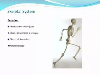
Skeletal System. Function : Protection of vital organs Muscle attachment & leverage Blood cell formation Mineral storage. Types of Skeletons. Axial (green) 80 bones skull, vertebral column, ribs, & sternum Appendicular (purple) 126 bones

Skeletal System
E N D
Presentation Transcript
Skeletal System Function : Protection of vital organs Muscle attachment & leverage Blood cell formation Mineral storage
Types of Skeletons Axial (green) 80 bones skull, vertebral column, ribs, & sternum Appendicular (purple) 126 bones Upper extremities {includes clavicles & scapula} Lower extremities { includes pelvis}
Bone Types Lippert p. 15, Table 2-2 • Long • length > width • Short • more equal dimensions • usually articulate w/ more than one bone
Flat:** broad, curved surfaces** site for red blood cell formation
Sesamoid: ** located where tendons cross a joint protection from excessive wear mechanical advantage
Bone Anatomy Epiphysis distal & proximal wider than shaft cancellous bone Epiphyseal Plate cartilaginous in growing bone Metaphysis flared ends of diaphysis cancellous bone supports epiphysis Diaphysis shaft compact bone
Bone Anatomy Cont’d Medullary Canal center of diaphysis hollow → decreases weight contains marrow, passage for nutrient arteries Endosteum lines medullary canal
Bone Anatomy Cont’d Periosteum thin, fibrous membrane covers all bone except articular surfaces contains nerves & blood vessels attachment point for tendons & ligaments
Bone Structure Overview Periosteum = Superficial layer Compact bone = Middle Cancellous bone = Deep
Bone Structure Compact / Cortical Bone: • hard / dense • weight bearing • Osteon : - structural unit of compact bone - “weight bearing pillars” • Lamella : - layered, hollow tubes - collagen of each layered tube runs in opposite directions • Central Canal : - runs vertically through center - contain nerves & blood vessels • Perforating Canal : - runs horizontally - connects blood & nerve supply periosteum → central canal of osteon
Bone Structure cont’d Cancellous/ Trabecular Bone: • “little beams” • no osteons present • Lamella = hollow tubes • irregularly arranged • surrounded by endosteum • filled w/ marrow • bone weight • shock absorption • found at articular ends of bones
Endochondral ossification Bone Development formation of long bones formed from cartilage Steps: - Development of primary ossification center - bone collar formation - calcification - cancellous bone formation - formation of medullary cavity - development of secondary ossification center
Intramembranous ossification • formed from mesenchyme tissue • occurs during formation of the flat bones of the skull • Steps: Development of ossification center → formation of bone matrix→ formation of trabeculae & periosteum→ formation of bone collar & red marrow
Joint Types • Fibrous : thin layer of fibrous periosteum between 2 bones, no cavity 1. Synarthrosis : ( suture jt.) ; no motion ; Ex. Skull 2. Syndesmosis: (ligamentous jt.); minimal motion- depends on length of connecting fibers; Ex. distal tibiofibular jt. distal radioulnar jt. 3. Gomphosis: (peg-in-socket); no motion; Ex. Tooth in socket of mandible/maxilla • Cartilaginous (Amphiarthrosis):bones united by cartilage, limited motion (bending ,twisting, compression) 1. Synchondrosis: hyaline cartilage; Ex. ephipyseal plate (children), 1st sternocostal jt. 2. Symphysis : fibrocartilage ; shock absorber; Ex. intervertebral discs, pubic symphysis
Joint Types cont’d Synovial (Diarthrosis) : fluid filled jt. cavity free motion w/ decreased stability Stability determined by : 1.) Shape of articular surface 2.) Number/position of ligaments 3.) Muscles & tendons crossing the jt. {Lippert p. 19}
Synovial Joints Plane: gliding motion Example : * inter tarsal * intercarpal
Synovial Joints Pivot: rotation Example: * proximal radioulnar jt. * atlantoaxial jt.
Synovial Joints Hinge:flexion/extension Example: * elbow * knee
Synovial Joints Condyloid: flexion/extension abduction/adduction Example: * wrist * metacarpalphalangeal jt.( MP)
Synovial Joints Saddle : flexion/extension abduction/ adduction rotation (accessory) Example: * carpometacarpal jt. (thumb)
Synovial Joints Ball & Socket: flexion/ extension abduction/ adduction rotation Example: * hip * glenohumeral jt. (GH)
Synovial Jt. Structure • Ligaments : • bone approximation • prevent excessive motion; stability • attachment for cartilage, fascia, & muscle • Capsule : • surrounds joint • protects articular surfaces • 2 layers : -- Outer = fibrous; reinforced by capsular ligaments -- Inner = synovial membrane • sensory nerve ending = proprioception • rich supply of blood vessels = nutrition
Synovial Jt. Structure cont’d Joint Cavity: “free space”
Synovial Jt. Structure cont’d • Hyaline Cartilage (articular) : • provides smooth articulating surface • no blood or nerve supply → unable to repair itself if damaged • Fibrocartilage : • shock absorption in weight bearing joints • *knee – menisci : improve stability vs. shock absorption • *intervertebral discs : shock absorption • *sternoclavicular jt. : shock absorption betwn. clavicle & sternum
Synovial Jt. Structure cont’d • Synovial Membrane : • inner layer • thick & vascular • secretes synovial fluid • Synovial Fluid: • found in jt. cavity & w/in articular cartilage • primarily from filtration of blood through capillaries of synovial membrane • clear, viscous consistency due to hyaluronic acid content • becomes less viscous w/ ↑ jt. motion / temp. • reduces friction • shock absorption • Nutrition: ** weight bearing → jt. compression → forcing synovial fluid out of cartilage → compression removed → synovial fluid seeps back into cartilage ( Weeping Lubrication)


