Download
1 / 99
1.02k likes | 1.91k Views
Lymph nodes and vessels. David Kachlík Martin Špaček Anne LeRoy. Lymph system. lymph ( lympha ) composition similar to plasma woth low protein content chylus ( chylos = gr. juice) – intestinal lymph, milky - chylomicrones ) lymph vessel ( vas lymphoideum ) lymph organs

E N D
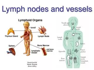
Lymph nodes and vessels David Kachlík Martin Špaček Anne LeRoy
Lymph system • lymph (lympha) • composition similar to plasma woth low protein content • chylus (chylos = gr. juice) – intestinal lymph, milky - chylomicrones) • lymph vessel (vas lymphoideum) • lymph organs • all lymph is drained into veins (into angulus venosus) • missing in some organs (cartilage, cornea, bone marrow, placenta) or replaced with other fluid (brain, eyeball, inner ear…)
Lymph system - function • blood filtration and „purification“ via lymph nodes – immune function • return of extravascular fluid into blood circulation • transport of proteins and other large particles into systemic circulation • „education“(maturation) and multiplication of lymphocytes • transport of lipids in chylomicrones from small intestine into systemic circulation • daily production = cca 60 ml / kg
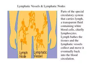
Lymph capillaries • begin as cul-de-sac • fluid flows only inside • plus: viruses, bacteria, tumour cells • collect superfluous tissue fluid • no lamina basalis • regularly one-way valves • parallel to venules • collect into lymphatic vessels
Lymph capillaries within villi • villi of small intestine • special arrangement • lymph capillary in villus axis • „lacteals“ • lipid absorption (chylomicrones)
Lymph vessels • thin-walled vessel (thinner than in veins) • interrupted lamina basalis • endothelium without pores and zonulae occludentes • anchoring filaments (enlarged openings between cells during swelling of surrounding tissue) • larger amount of vessels • lymph nodes put in course of lymph vessels • lymph trunks: rather thick tunica media but thin tunica externa
Lymph vessels general arrangement • lymph capillaries lymph vessels • limbs (collectors):superficial deep vessels • organs: plexuses (subcapsular + deep) lymph trunks lymph ducts • lymph nodes put in vessels
Lymph ducts 2 lymph ducts • irregular division of body (drainage areas) • ductus thoracicus • pars abdominalis • cisterna chyli Pecqueti (50%) • pars thoracia • pars cervicalis • arcus d.t. • ductus lymphaticus dexter 9 lymph trunks
cirrhosis Ductus thoracicus et angulus venosus
CISTERNA CHYLI cholagiography
Lymph organs = acquired (adaptive, specific) immunity • primary (central) lymph organs • thymus • bone marrow = medulla ossium • bursa Fabricii – birds function: cell production de novo, differentiation and maturation of immunocompetent cells • secondary (peripheral) lymph organs • spleen = splen (lien) • lymph nodes = nodi lymphoidei • tonsils = tonsillae • lymph nodules = noduli lymphoidei function: contact with antigen-presenting cells, production of mature lymphocytes Světla blikat
Lymph nodes • lymph filtration • antigen presentation • activation, differentiation and proliferation (production of mature B- and T-lymphocytes) • lymphocytes, macrophages, plasmatic cells
Lymph node • nodus lymphoideus (nodus lymphaticus; lymphonodus) • cca 500 in body • ø 1-25 mm • swelling in inflammation (painful) • metastasis catching (inpainful swelling) • „lymphadenopathy“
Lymph node • capsula trabeculae • hilum • cortex (+ paracortex) • reticulin network • medulla • convex part - „afferent“ (more afferent lymph vessels) • concave hilum - „efferent“ (one efferent lymph vessel + blood vessels)
Lymph node - cortex • outer cortical zone (cortex) • sinus subcapsularis • lymph nodules (noduli lymphoidei) • sinus internodulares • B-lymphocytes • inner cortical zone(paracortex) • T-lymcytes (CD4, CD8) • dendritic cells • no lymph nodules • venulae altoendotheliales • high endothelium
Lymph node – cortex: cells and zones noduli lymphoidei • B-lymphocytes • follicular dendritic cells (FDC) • primary nodule • small „naive“B-lymphocytes • secondary nodule • germinal center: antigen-stimulated B-lymphocytes (large, less deeply staining, more rapidly dividing) • dark zone: centroblasts • light zone: centrocytes • other cells: T-lymphocytes (help maturation of B-lymphocytes), macrophages (phagocyte apoptotic cells) • mantle zone • produced as surrounding cells are marginalized by rapidly growing germinal centre • naive B-lymphocytes, TH-lymphocytes, FDC, macrophages
Lymph node - medulla • chordae medullares (medullar cords) • processes of cortical lymphoid tissue • many B-lymphocytes and plasmatic cells • dendritic cells (antigen-presenting cells) • sinus lymphaticus medullaris • lined with reticular cells and macrophages • reticular fibers cross sinuses – web
Lymph node – blood and lymph flow vessels: „naive“ lymphocytes from primary lymphoid organs vasa lymphatica afferentia: lymph with antigenes and antigen-presenting cells (APC) sinus subcapsularis sinus internodulares (radiate from capsule into medulla) sinus lymphaticus medullaris vas lymphaticum efferens vas lymphaticum efferens: mature effector cells
TONSIL lymph nodule germinal center CD38 B-lymphocytes (red) mantle zone: IgD+ naive B-lymphocytes (green) activated B-lymphocytes, transferrin receptor (CD 71) – positive cells of various cellular lines (blue)
Lymph nodule – cell morphology • small naive B-lymphocytes • small round dark blue dots • round nucleus, clumped chromatin, small or absent nucleolus • imunoblasts • large cell • large nucleus and one centrally located nucleolus • centroblasts • large cell with vesicular chromatin • 2-3 nucleoli, rare cytoplasm • centrocytes • moderately large cell • cleaved irregular nucleus and condensed chromatin
Recirculation of lymphocytes 1. • lymphocytes leave lymph nodes and together with lymph enter venous blood • during return („homing“) inform other lymphoid organs on activation of certain satellite lymph node organism preparation to generalized immune response • organ specificity during „homing“ is assured with chemotactic cytokines and combination of adhesive molecules • on lymphocytes and on specific high endothelium of postcapillary venules(venulae altoendotheliales) in paracortex
Recirculation of lymphocytes 2. based on specific adhesive molecules, lymphocytes attach to venular endothelium and squeeze through endothelial cells lymphocytes adhere to luminal surface of endothelial cells thanks to microvilli
Recirculation of lymphocytes 3. • B-lymphocytes then migrate into outer cortical zone (cortex) • T-lymphocytes rest mainly in paracortex
Lymph nodes – distribution • tributary regions • regional lymph nodes • sentinel lymph node • inflammation – painful swelling • tumour – unpainful swelling • lymphedema (elephantiasis) TNM classification
TNM classification • tumor • nodus • metastasis • contact (direct spreading) • lymphogenous • haematogenous
Head • n.l. occipitales • rubella • toxoplasmosis • n.l. mastoidei • n.l. parotidei • n.l. faciales
Neck n.l. cervicales • anteriores • laterales n.l.c. profundi n.l. jugulodigastricus n.l. juguloomohyoideus n.l.c. superficiales • chain along v. jugularis int. (= n.l. cervicales laterales profundi) • chain along n. XI (n.l. accessorii) • n.l. supraclaviculares
Neck • n.l. submentales • n.l. submandibulares • n.l. pretracheales • n.l. paratracheales • n.l. retropharyngei • Rouvière‘s node • childern, mesotitis
Clinical classification of cervical lymph nodes 6 quadrants Robbins, 2001
6 levels (quadrants) of cervical lymph nodes Ia: lower lip, mouth, anterior 1/3 of the tongue, anterior 1/3 of the mandibular alveolar ridge Ib: mouth, nasal cavities, skin and soft tissues of the ipsilateral midface II:mouth, nasal cavities, nasopharynx, oropharynx, hypopharynx, larynx, parotid glands III: mouth, nasopharynx, oropharynx, hypopharynx, larynx IV: hypopharynx, larynx, cervical esophagus V:nasopharynx + oropharynx VI: thyroid gland, larynx, cervical esophagus
Virchow-Troisier • n.l. supraclaviculares sinistri • enlarged in: • tumor of stomach (70%) • tumor of left breast • tumor of lung • tumor of large intestine • tumor of neck on the left side • primary lymphoma
Tongue • 4 directions • n.l. submentales (apex) • n.l. submandibulares (corpus margines) • n.l. cervicales profundi (corpus center + radix) • contralaterally across midline
Axilla up to 40 5 groups • apicales • centrales • humerales • subscapulares • pectorales
Mammary gland • n.l. pectoralis Sorgiusi • 2nd/3th tooth of m. serratus ant. • sentinel lymph node • n.l. infraclaviculares supraclaviculares • n.l. parasternales n.l. mediastinales ant. • across midline n.l. axillares contralaterales
Mammary gland • 4 quadrants • 2 superficial plexuses: • plexus dermalis • plexus subareolaris Sappeyi • drainage into n.l. axillares • 2 deep plexuses: • plexus fascialis • plexus glandularis • drainage into superficial plexuses and other lymph nodes • 80 % of lymph into n.l. axillares