Download
1 / 92
1.14k likes | 4.23k Views
Pathology of Lymph Nodes. Norman Levy, MD. Big Picture. As with other organs, lymph nodes, and more globally, the immune system, can be the site of infectious, immune and neoplastic disease, the latter either primary or metastatic

E N D
Pathology of Lymph Nodes Norman Levy, MD
Big Picture • As with other organs, lymph nodes, and more globally, the immune system, can be the site of infectious, immune and neoplastic disease, the latter either primary or metastatic • The clinical manifestations of diseases of the lymph nodes are: • Local enlargement, tender on nontender, +/_ • Compression of adjacent structures +/_ • Release of cytokines producing "systemic" symptoms of fever, weight loss and night sweats • Infectious organisms can stimulate the same acute, chronic or granulomatous reactions in the draining lymph nodes as they characteristically stimulate at other sites
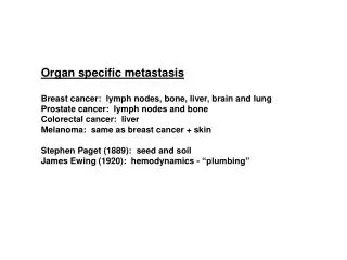
Big Picture 2 • Several types of immune stimuli can cause "reactive" enlargement of the entire lymph node, or selective expansion of cortical, paracortical or medullary regions • Metastatic tumors spread to the lymph nodes primarily via lymphatic drainage from adjacent solid organs • Primary neoplasms of the lymph nodes are all malignant • They are divided into malignant non-Hodgkin's lymphomas (NHL), and Hodgkin lymphoma
Big Picture 3 • NHL's are more common, and can be simply divided into indolent, or slow growing types, and aggressive types • Malignant lymphomas represent clonal malignancies in which mutational events have caused the majority of progeny cells to freeze at a single stage of normal lymphocyte differentiation • Lymphomas frozen at a stage associated with high replication --> aggressive lymphomas; • Lymphomas frozen at stages associated with recirculation or final function --> indolent lymphomas
Big Picture 4 • The diagnosis of malignant lymphomas is based on the microscopic recognition of the dominant cytologic cell type, supplemented by immunologic and molecular techniques • The treatment and prognosis of lymphomas are based on • The dominant cell type (and it's inherent biologic behavior), • The extent of spread (Stage) • The underlying health of the patient • All of the previous statements are complicated by the fact that indolent lymphomas can further mutate and transform to aggressive types
Big Picture 5 • Hodgkin lymphoma is a less common nodal disease whose diagnosis is based on the detection of a characteristic cell, the Reed Sternberg cell, in the appropriate histologic setting • There are several (five) histologic subtypes, but prognosis is based primarily on extent of disease • Hodgkin lymphoma is a more curable disease than non-Hodgkin lymphomas • Now watch me confuse this relatively straightforward information with the details.
Overview of the lymphoid immune system • Lymphocytes evolve from pluripotent stem cells --> two major functional cell types: • B lymphocytes, comprising the humoral immune --> production of antibodies • T lymphocytes, comprising the cellular immune system, --> • Direct killing of foreign or intracellularly infected cells, cytotoxic T cells • Fine control of the immune response through the secretion of cytokines, helper and suppressor T cells.
Anatomicalorganization • The anatomic organization of the lymphoid immune system divided into two major functional regions: • The primary immune organs, sites of initial maturation --> immune competent cells: • B cells- bone marrow • T cells- thymus • The secondary immune organs, sites of antigen driven replication and differentiation into committed effector cells • Lymph nodes • Spleen • Mucosal Associated Lymphoid System (MALT)- lymphoid cells lining the respiratory and gastrointestinal tracts • Everywhere else • The lymph nodes, in their totality, represent the largest secondary organ, and the major site of lymphoid pathology
Lymph node anatomy • To recognize lymph node pathology, one has to be familiar with normal lymph node anatomy and cytology
Lymph node variation • Lymph node histology is dynamic: follicles • In the absence of immune stimulation, primary follicles • In the presence of immune stimulation, secondary follicles or germinal centers
After initial maturation in the primary immune organs, "virgin" B and T lymphocytes --> peripheral blood --> home to specific sites within the lymph node (and the other secondary organs), The sites of B cell homing include: The primary and secondary follicles of cortex-the sites of antigen presentation proliferation and differentiation in response to same The medullary cords -->plasma cells aggregate--> release their immunoglobulins into the efferent lymph The site of T cell homing is the paracortex The separation of B and T lymphocytes not absolute, Both cell types present throughout lymph node, necessary for coordinated lymphoid immune response. Lymphocyte homing
Lymphocyte recirculation • Normal lymphocytes recirculate, passing from blood --> lymph nodes --> efferent lymphatics • Allows constant surveillance for the presence of the antigen for which the lymphocyte has a unique and specific receptor on it's surface. • If antigen not present, lymphocytes leave the node and recirculate • Virgin lymphocytes have a finite lifespan, numbered in weeks, unless they come in contact with antigen
Cytology of the lymph node • The normal or reactive lymph node is thus a dynamic organ • Composed of • Transient B and T lymphocytes • Antigen processing and presenting cells • Replicating B and T lymphocytes (in response to antigen) • Persistent and transient final effector cells • Macrophages • Some of these functional subgroups are cytologically unique, others cytologically indistinguishable • The ultimate microscopic impression, with practice, is one of cytologic heterogeneity, and histologic organization
Small lymphocytes Small round dark blue dots. Round nucleus, clumped chromatin, small or absent nucleolus. The dullest looking cells hiding the greatest level of functional heterogeneity. Can be T or B cell, virgin (unexposed to antigen) or differentiated effector/memory cell. Most likely lineage, B or T, guessed by location within the node, but lineage and state of differentiation must be confirmed by immunologic/molecular techniques Locations: B cells- primary follicles, mantle zone of secondary follicles, medullary cords T cells- paracortex, minor population within germinal center. Kinetically, clumped chromatin tells us that the cell is not proliferating- not activated to enter the cell cycle and replicate Cell types I
Replicating and post-replicating B cells Noncleaved cells, small and large Replicating populations- expanding antigen responsive cells. Round nuclei but larger than resting small lymphocyte Open or vesicular chromatin Recognizable nucleoli. Nucleus clear -->genetic material unwound for replication. Size, large or small compared nucleus of macrophage. Small cleaved cells- Nonreplicating population Post mitotic memory or plasma cell precursors Clumped chromatin Irregular folded and cleaved nuclear profiles Cell types 2:Follicular (germinal) center cells
Reactive germinal center MZ LZ DZ
Immunoblasts Replicating large cells found outside the germinal centers. May be of B or T cell type Have nuclear characteristics of replicating lymphocytes- Vesicular chromatin Nucleoli Accessory cells Antigen processing cells Interdigitating reticulin cells- T cell paracortex Dendritic reticulin cells- B cell germinal centers Process and present antigen to B and T lymphocytes Invisible in normal lymph node Macrophages (histiocytes)- Phagoctytic cells of lymph node Tingible body macrophages of germinal centers Medullary and subcapsular sinus macrophages- Abundant pale cytoplasm Oval nucleus, single small nucleolus Cytology of lymph node 3
Pathology of lymph nodes 1 • Infections • Reactive hyperplasias • Sarcoidosis • Metastatic tumors • Malignant lymphomas • Non-Hodgkin’s lymphoma-NHL • Hodgkin’s lymphoma
Pathology of lymph nodes 2 • Infections • Bacterial • Acute inflammation, abscess formation • Granulomatous, caseous and noncaseous • Diagnosis by culture, serologies, and/or special stains
Reactive hyperplasias • Exaggerations of normal histology. • Expansion of all regions or selective expansion • Some types characteristic of certain diseases, but most not • Follicular hyperplasia- increase in number and size of germinal centers, spread into paracortex, medullary areas • Collagen vascular diseases • Systemic toxoplasmosis • Syphillis • Interfollicular hyperplasia- paracortex • Skin diseases • Viral infections • Drug reactions • Sinus histiocytosis- expansion of the medullary sinus histiocytes- • Adjacent cancer • Infections
Malignant lymphomas (Non-Hodgkin's lymphomas-NHLs) • Malignancies of the lymphoid system which primarily manifest themselves outside the bone marrow, at the sites of normal lymphoid homing • Lymph nodes • Spleen • M.A.L.T. • Anywhere(Lymphomas outside lymph nodes and spleen are referred to as extranodal lymphomas) • Approximately 40, 000 cases per year, 20,000 deaths
Clinical presentation • Enlarging mass(es), typically painless, at sites of nodal tissue • Compression, infiltration of hollow organs • Pain, obstruction, perforation • Interference with normal organ function- • Solid organ infiltration- kidneys, liver, bone marrow • Systemic symptoms • Fever • Night sweats • Weight loss • If marrow infiltrated, can have leukemic component
NHL 2 • Composed of cells that have lost the ability to pursue the full range of lymphoid differentiation, and are frozen at a single stage of the normal maturation/differentiation sequence • Recapitulate the biology and immunophenotype of normal cell counterpart • Several cytologically and immunologically recognizable stages of normal lymphoid maturation --> several subtypes of lymphoma • Clonal malignancies, derived from a single cell that has undergone a malignant transformation, mutation • Best initially conceptualized as two major clinical types • Indolent lymphomas • Aggressive lymphomas
NHL 3 Indolent lymphomas • Lymphomas frozen at stages not normally replicating, but may be circulating • Diseases of slow accumulation, due to defective apoptosis • Often widespread at diagnosis • Prolonged natural history, median survivals >5 years • Will usually respond to chemo- or radiation therapy • Will usually relapse, but respond to same or alternative tx • Currently incurable unless • Localized disease or • Marrow ablation with some type of stem cell transplant • Classification of indolent lymphomas- later
Aggressive lymphomas • Lymphomas frozen at stages characterized by replication and accelerated growth • Diseases of defective cell cycle control • More often localized at presentation than indolent lymphomas • More often extranodal • Shorter natural history; median survival </= 2 years • Require more aggressive therapy to achieve "clinical remission"- disappearance of all detectable disease • Despite short natural history, curable disease in some with aggressive therapy • Approximately 30-40% of adults • 50-80% children • All childhood lymphomas of this type
Classification of lymphomas • Subtyping or classification within the two groupings necessary, because different subtypes have • Distinct clinical presentations • Can require different therapy • Have differing prognoses, reflecting different mechanisms of molecular pathogenesis. • Unfortunately, rarely unanimous acceptance of any one classification scheme. • Intermittent upgrading of classification, with new terminology, reflecting new information and classifier bias • Classification often lags behind advances in immunology, research pathology • Final result: • Difficult area to teach • Difficult to remember • Job security for me
From 1982-1994, the classification used in the United States Based on: The observed clinical history of 1200 patients classified according to the terminology to right Microsopic examination alone, utilizing Loss of normal nodal architecture The dominant cytologic cell type observed under the microscope Presence or absence of "follicularity" - mimicking of normal lymphoid follicle formation Low grade ML, small lymphocytic ML, follicular small cleaved cell ML, follicular, mixed small and large cell Intermediate grade: ML, follicular, large cell ML, diffuse, small cleaved cell ML, diffuse, mixed small and large cell ML, diffuse, large cell High grade ML, immunoblastic ML, lymphoblastic ML, small non-cleaved cell (Burkitt's vs non-Burkitt's) Miscellaneous (mycosis fungoides, true histiocytic, etc.) WorkingFormulation for Clinical Usage
Working Formulation • Divided into three "grades" of lymphoma- low, intermediate and high. As stated above, • Low grade = indolent • Intermediate and high = aggressive • Limitations • Purely morphologic classification mixed T and B cell lymphomas together • Lumped distinct subtypes of B cell lymphomas together • Obscured the biologic, clinical and therapeutic differences • Distorted interpretation of clinical trials
R.E.A.L./W.H.O. Classification • WF replaced in 1994 by the Revised European American Lymphoma (REAL) classification, now being modified by the World Health Organization (WHO) • REAL/WHO is a "disease” oriented rather than purely morphology oriented classification, based on: • Cell lineage: B v T v NK v Histiocytic • Stage of maturation of the presumed normal counterpart. • Includes immunologic and molecular criteria in addition to purely morphologic criteria of WF • Each disease entity may have differing grades of aggressiveness • Greatly expanded the list of entities; includes leukemias of lymphoid origin • Made teaching to medical students (and in fact all physicians) even more difficult than WF • REAL contained a number of “provisional entities” which have been clarified in the upcoming W.H.O. revision.
REAL/WHO classification- backbone • B cell neoplasms • Precursor B cells-related to acute leukemia • Peripheral B cell lymphomas- the majority of B cell lymphomas • T cell and Natural Killer cell neoplasms • Precursor T cells • Peripheral T cell and NK neoplasms • Hodgkin’s lymphoma
Indolent Small lymphocytic lymphoma/CLL Follicular lymphoma, Grades 1/2 Extranodal Marginal zone lymphoma of MALT type Nodal marginal zone lymphoma Splenic marginal zone lymphoma Hairy cell leukemia Lymphoplasmacytic lymphoma Plasma cell myeloma Plasmacytoma Cutaneous T cell lymphoma Cutaneous CD30+ anaplastic large cell lymphoma Aggressive Prolymphocytic leukemia Large B cell lymphoma Burkitt lymphoma Mantle cell lymphoma Anaplastic large cell lymphoma All peripheral T cell lymphomas Indolent versus aggressive Divides B and T
B cell neoplasms- Precursor B • Precursor B cell lymphoblastic leukemia/lymphoma • Frozen at lymphoblast cell stage of antigen independent B cell differentiation- normally restricted to bone marrow • Usually present as acute leukemia +/- lymph node involvement • Can initially present as node or skin disease, with later progression to bone marrow • Treated as acute leukemia • 80% cure rate in children • 20-30% in adults because of "bad" cytogenetics: frequent presence of Philadelphia chromosome t(9;22)
Peripheral B-cell lymphomas • Lymphomas frozen at various stages of antigen dependent B cell maturation and differentiation
Peripheral B-cell neoplasms • Frozen at various stages of antigen dependent B cell maturation and differentiation • Small lymphocytic/CLL- the virgin B cell fresh from the marrow • Prolymphocytic leukemia- a more clinically aggressive variant of above • Lymphoplasmacytic lymphoma- the primary immune response • Mantle cell lymphoma- the mantle region surrounding the follicle • Follicular lymphoma- the follicle- grades 1-3 • Extranodal marginal zone lymphoma- cells at the periphery of the follicle in extranodal sites of lymphoid tissue- Mucosal Associated Lymphoid tissue- such as G.I. tract • Nodal marginal zone lymphoma • Splenic marginal zone lymphoma- immunologically distinct • Hairy cell leukemia- pre-plasma cell • Diffuse large B-cell lymphoma- this breaks the ideal of specific cell stage but all represent lymphomas with high replication rate • Burkitt lymphoma- very aggressive • Plasma cell myeloma- diffuse bone marrow proliferation of plasma cells • Plasmacytoma- solitary focus of monoclonal plasma cells, with variable risk of progression to myeloma, depending on site
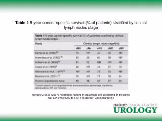
Clinical Most common type of indolent lymphoma in US; second most common type lymphoma overall Disease of adults >40 (median age 59) Usually widely disseminated at diagnosis, incl. bone marrow Will respond to “gentle chemotherapy” but will relapse Incurable short of bone marrow transplant unless rare limited disease Overall 5 yr survival 72% Over time, additional mutations --> progression (“transformation”) to large cell lymphoma --> aggressive clinical course Although Gr.1 is most common presentation, some patients present with predominance of large cells within follicles -->more aggressive clinical course Pathogenesis: Due to t(14;18)(q32, q21) Upregulates expression of an anti-apoptotic protein Bcl2 Immortalizes lymphoma cells Example Indolent Lymphoma:Follicular lymphoma Grade I
Follicular lymphoma Grade I • Pathology/diagnosis • Benign equivalent: small cleaved cell of germinal center • Clumped chromatin and infrequent nucleolus like small lymphocyte • Irregular nuclear profile, with nuclear folds or "cleavages" • Retain follicular structure, but monotonous accumulation of single cell type • Characteristic immunophenotype: • Positive:Monoclonal light chain, CD19, CD10, Bcl2 • Negative: CD5, Cyclin D1/Bcl1 • Can also detect translocation by cytogenetics and/or polymerase chain reaction
Examples: aggressive B cell lymphoma-Diffuse large B cell lymphoma • Clinical • Most common lymphoma- 30% NHL • Disease of adults and children, but median age 64 • Limited versus widespread disease ~1:1 • Presents with rapidly enlarging masses • Approximately 40% curable with aggressive chemotherapy/ stem cell transplant • Partially predictable by International Prognostic Index (later) • Pathogenesis • Not as clearly defined as previous examples- several cytogenetic abnormalities associated with large cell lymphoma, but no defining one
Diffuse Large B cell lymphoma • Pathology • Benign equivalent- large replicating B cells of germinal center and paracortex • Diffuse infiltration of lymph node • Often necrosis; increased mitotic rate • Cytology: Oval or cleaved nucleus with vesicular chromatin and 1-3 nucleolus • Nucleus larger than that of reactive macrophage • Several cytologic subtypes initially felt to have differing clinical behavior. • Yielded division into intermediate versus high grade types- now not felt valid or significant without immunologic/molecular evidence • Immunophenotype characterized by monoclonal light chain, CD19 expression,with variable expression of other B cell associated antigens
Clinical 3% lymphomas Disease of adults and children- median age 31 Initially recognized in Africa by Thomas Burkitt Association with Epstein Barr virus infection Localization in jaw In US, usually presents in ileocecal region of children 1/3 of all childhood lymphomas Earlier eras, very aggressive and rapidly fatal Now, ~70-80% children curable 40% of adults Pathogenesis: t(8;14), producing upregulation of myc oncogene, a cell cycle regulation gene Burkitt's lymphoma
Burkitt's lymphoma • Pathology • Benign equivalent is replicating small noncleaved cell of germinal center: • Diffuse infiltration of lymph node • Very high mitotic rate, lot of ineffective proliferation; • Attracts macrophages to phagocytize> starry sky pattern at low power • Cytology: round nucleus, smaller than that of reactive macrophage • Vesicular chromatin and 2-5 nucleoli • Immunophenotype: • Positive: Monoclonal light chain, CD19, CD10 • Negative: CD5
Mantle cell lymphoma • Clinical • 6% lymphomas • Disease of adults (median age 63) • Usually widely disseminated • Poor response to all attempted therapies, • ? curable with transplant • 5yr survival 27% • Pathogenesis • Due to t(11;14) • Upregulates Bcl1 (cyclin D1), a cell cycle regulator
Mantle cell lymphoma • Pathology/Diagnosis • Benign equivalent is lymphocyte of inner mantle zone • Cytology similar to cleaved cell, but nuclear irregularities not as prominent • Nodal infiltration diffuse, vaguely nodular or "mantle zone" around residual benign follicles • Large cell progression infrequent • Immunophenotype: • Positive: monoclonal light chain, CD19, CD5, Bcl1 (and Bcl2) • Negative CD10, CD23
Follicular lymphoma Mantle cell lymphoma CyclinD1 Bcl2