Download
1 / 14
180 likes | 343 Views
Lymph nodes are small, bean-shaped structures crucial to the lymphatic system, located at the junctions of lymphatic networks. They play vital roles in filtering lymph, facilitating immune responses, and harboring memory cells for quicker responses to antigens. Each lymph node comprises an outer cortex, inner medulla, and is covered by a fibrous capsule. The spleen, with its thick capsule and trabeculae, consists of white pulp (lymphatic nodules) and red pulp (blood sinuses). Understanding these structures aids in comprehending their important functions in immunity and blood filtration.

E N D
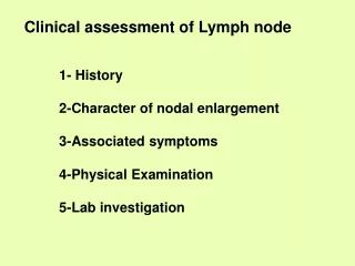
LYMPH NODE • Small bean shaped structures • Found at junctions of lymphatic networks. • Covered by a fibrous capsule. • Has a subcapsular sinus connected to subtrabecular, and medullary sinus • Afferent lymph vessels open into subcapsular sinus • Efferent lymphatics leave the hilus. • Has outer cortex and inner medulla.
FUNCTIONS OF LYMPH NODE- 1)Filters the lymph and macrophages carry out phagocytosis. 2)Concentration on antigens and their presentation to the lymphocytes. 3)Cell mediated and humoral immunity. 4)Memory cells-more rapid response to antigen when it is next encountered.
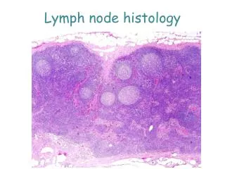
MICROSCOPIC FEATURES • Covered by capsule which sends in trabeculae. • Outer CORTEX has rounded lymphoid nodules made up of lymphocytes PRIMARY NODULE • Some nodules show GERMINAL CENTRES SECONDARY NODULE • Inner MEDULLA contains lymphocytes arranged in cords called medullary cords between medullary sinuses.
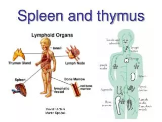
SPLEEN Vascular friable organ Capsule thick & trabeculae thick. Parenchyma divided into white pulp& red pulp. White pulp is lymphatic nodule with eccentric arteriole. Red pulp contains blood sinuses with RBC. In between blood sinuses and rbc, lymphocytes are arranged in cords called Billroth cords.
SPLENIC CIRCULATION • OPEN CIRCULATION • CLOSED CIRCULATION
OPEN CIRCULATION- • The capillaries end by opening into the red pulp from where the blood enters the sinusoids through their walls.
CLOSED CIRCULATION- • The capillaries are continuous with the venous sinusoids in the red pulp and sinusoids join together to form splenic veins.
LYMPH NODE Capsule & subcapsular sinus. Cortex with lymphatic nodules with germinal centres. Medulla with cords &sinuses. SPLEEN Thick capsule & trabeculae. White pulp & red pulp. White pulp contains eccentric arteriole. LYMPH NODE & SPLEEN (main features)