Download

1 / 38
380 likes | 486 Views
The lymphatic system comprises a network of lymphatic vessels and various lymphoid tissues and organs throughout the body. It serves to return interstitial fluid and leaked plasma proteins to the bloodstream; once inside the vessels, this fluid is termed lymph. Key components include highly permeable lymphatic capillaries, collecting vessels, and trunks. The system plays a vital role in immune function, facilitating the transport of lymphocytes, macrophages, and filtering pathogens and debris to maintain bodily health.

E N D
20 The Lymphatic System
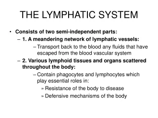
Lymphatic System: Overview • Consists of two semi-independent parts: • A network of lymphatic vessels • Lymphoid tissues and organs scattered throughout the body • Lymphatic vessels return interstitial fluid (fluid in the tissues) and leaked plasma proteins back into the bloodstream • Once interstitial fluid has entered lymphatic vessels, it is called lymph
Lymphatic System: Overview Figure 20.2a
Lymphatic System: Overview Figure 20.1a
Lymphatic Vessels • One-way system, lymph flows toward the heart • Lymph vessels include: • Microscopic, permeable, blind-ended capillaries • Lymphatic collecting vessels • Trunks and ducts
Lymphatic Capillaries • Similar to blood capillaries, with modifications: • Very permeable • Loosely joined endothelial minivalves • Withstand interstitial pressure and remain open • The minivalves function as one-way gates that: • Allow interstitial fluid to enter lymph capillaries • Prevent lymph from escaping out of the capillaries
Lymphatic Capillaries Figure 20.1b
Lymphatic Capillaries • During inflammation, lymph capillaries can absorb: • Cell debris • Pathogens • Cancer cells • Lacteals – specialized lymph capillaries present in lining of the small intestines • Absorb digested fat and deliver chyle(fatty lymph) to the bloodstream
Lymphatic Collecting Vessels • Have the same three tunics as veins • Have thinner walls, with more internal valves • Anastomose(branch off) more frequently • Collecting vessels in the skin travel with superficial veins • Deep vessels travel with arteries
Lymphatic Trunks • Lymphatic trunks are formed by the union of the largest collecting ducts • Lymph is delivered into one of two large trunks • Right lymphatic duct – drains the right upper arm and the right side of the head and thorax • Thoracic duct – arises from the cisternachyli and drains the rest of the body
Lymphatic Trunks Figure 20.2b
Return of Lymph to Heart Figure 20.2a
Lymph Transport • The lymphatic system lacks a pumping organ • Vessels operate under low pressure • Uses the same methods as veins to propel lymph: • Pulsations of nearby arteries • Contractions of smooth muscle in the vessel walls • Contractions of skeletal muscle
Major Tissue Type: Reticular Connective Tissue • Network of reticular fibers. Weblike. • Loosely bound together to support lymphocytes and macrophages
Lymphoid Cells • Lymphocytes are the main cells involved in the immune response • Two main varieties: • T cells • B cells • Both are produced in the red bone marrow – so it is classified as a primary lymphatic organ. • B cells mature in the red bone marrow. • T cells must travel to the thymus gland to mature; thus the name “T”
Lymphocytes • T cells • Manage the immune response • Directly attack and destroy virus - infected cells and tumor cells • B cells • Produce antibodies which circulate in the blood and attack foreign substances (antigens)
Macrophages • Macrophages – big “eaters” • When monocytes leave the blood stream and enter tissues they transform into macrophages and “eat up” viruses, bacteria, especially in chronic infections like T.B.
EDEMA • Swelling caused by abnormal amount of interstitial (between cells) fluid. • Common in lower extremities when right side of the heart is damaged. • Common in lungs when left side of the heart is damaged.
Lymph Nodes • Principal lymphoid organs of the body • Embedded in connective tissue and clustered along lymphatic vessels • Aggregations of these nodes occur near the body surface in inguinal, axillary, and cervical regions of the body Figure 20.4a
Lymph Nodes • Two basic functions: • Filtration – macrophages destroy microorganisms and debris • ***Only part of the lymphatic system that filters lymph *** • Immune system activation – monitors for antigens and mounts an attack against them
Structure of a Lymph Node Bean shaped and surrounded by a fibrous capsule Figure 20.4a, b
Circulation in the Lymph Nodes • Lymph enters via afferent lymphatic vessels • It then enters a large sinus and travels into smaller sinuses • It meanders through these sinuses and exits the node at the hilus via efferent vessels • Because there are fewer efferent vessels, lymph flow slows down in the node • This allows lymphocytes and macrophages time to carry out protective functions
Lymphoid Organs Figure 20.5
Other Lymphoid Organs • The spleen, thymus gland, and tonsils • Peyer’s patches and bits of lymphatic tissue scattered in connective tissue • All are composed of reticular connective tissue • All help protect the body
Spleen • Largest lymphoid organ, located on the left side of the abdominal cavity beneath the diaphragm • It is served by the splenic artery and vein, which enter and exit at the hilus • Functions: • Site of lymphocyte proliferation (in WHITE pulp) • Immune surveillance and response • Cleanses the blood
Additional Spleen Functions • Breaks down and stores products of RBCs for later reuse (in RED pulp) • Spleen macrophages salvage and store iron for later use by bone marrow • Site of fetal erythrocyte production (normally ceases after birth) • Stores blood platelets • Does NOT filter lymph
Structure of the Spleen Highly vascular Figure 20.6a, b
Thymus • A bilobedorgan located • in the inferior neck and extends into the mediastinum where it partially overlies the heart • Size of the thymus varies with age: • It increases in size and is most active during childhood • It stops growing during adolescence and then gradually atrophies • By this time, enough T lymphocytes have matured; vaccinations and antibodies formed from exposure to illnesses has occurred
Thymus • The thymus differs from other lymphoid organs in two important ways • Secretes hormones that cause T lymphocytes to mature, enabling them to recognize pathogens and initiate an immune response • It does not directly fight antigens • It is classified as a primary lymphatic organ.
Tonsils • Simplest lymphoid organs; form a ring of lymphatic tissue around the pharynx • Location: • Palatine tonsils – either side of the posterior end of the oral cavity • Lingual tonsils – lie at the base of the tongue • Pharyngeal tonsil – posterior wall of the nasopharynx (referred to as ADENOIDS if enlarged) • Tubal tonsils – surround the openings of the auditory tubes into the pharynx
Tonsils • Lymphoid tissue of tonsils contains follicles with germinal centers • Tonsil masses are not fully encapsulated • Epithelial tissue overlying tonsil masses invaginates (folds in on itself), forming blind-ended crypts • Crypts trap and destroy bacteria and particulate matter
Aggregates of Lymphoid Follicles • Peyer’s patches – isolated clusters of lymphoid tissue, similar to tonsils • Found in the wall of the distal portion of the small intestine • Similar structures are found in the appendix • Peyer’s patches and the appendix: • Destroy bacteria that entered the body via the mouth or anus, preventing them from penetrating the intestinal wall • Generate “memory” lymphocytes for long-term immunity
MALT • MALT – mucosa-associated lymphatic tissue • Protects passages that open to the EXTERIOR • Peyer’s patches, tonsils, and the appendix (digestive tract – mouth and anus) • Lymphoid nodules in the walls of the bronchi (respiratory tract – mouth and nose and ears) • Mucosal lining of the reproductive and urinary system ( urethra and vagina)
Breast Cancer/Lymph Nodes Axillary lymph nodes
Lymph Node Biopsy • Standard treatment for breast cancer was removal of the breast(s) and the axillary lymph nodes. • Now it is sometimes possible to biopsy the sentinel nodes (those closest to the tumor). • Dye is injected near the tumor and then sentinel nodes are identified if they are stained with dye. • If cancer has not spread, surgery is less invasive. • Sentinel node biopsy is used with other tumors, not just breast.
Post Surgery Care • When multiple regional lymph nodes are removed, the patient may experience side effects such as lymphedema (swelling caused by excess fluid build-up), numbness (nerve damage), infection and difficulty moving the affected body area. • Patients must avoid pressure (lifting heavy objects, having blood pressure taken, etc.) in affected area. • Wear an elastic stocking to reduce swelling, especially when flying.