Download
1 / 48
480 likes | 843 Views
Genitourinary Infections and Sexually Transmitted Diseases. 高雄長庚醫院婦產部 婦癌科 吳貞璇醫師. 1. Vulvar : sebaceous secretion, sweat, Bartholin, and Skene glands 2. Vagina : transudate from the vaginal wall; exfoliated vaginal and cervical cells; cervical

E N D
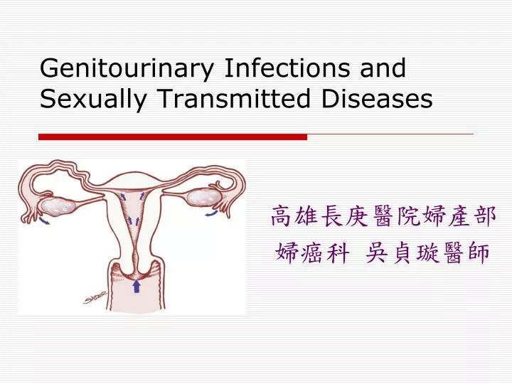
Genitourinary Infections and Sexually Transmitted Diseases 高雄長庚醫院婦產部 婦癌科 吳貞璇醫師
1. Vulvar: sebaceous secretion, sweat, Bartholin, and Skene glands 2. Vagina: transudate from the vaginal wall; exfoliated vaginal and cervical cells; cervical mucus; endometrial and oviductal fluids;and micro-organisms’ metabolic products --Increased during middle of the menstrual cycle The Normal Vagina Secretion
Vaginal epithelial cells • Affected by estrogen and progesterone: • Superficial cells: main cell type in reproductive age, predominate when estrogen stimulation. • Intermediate cells: predominate during the luteal phase because of stimulation by progesterone • Parabasal cells: in postmenopausal age
The normal vaginal flora • Mostly aerobic • 6 different species of bacteria, the most common of which is hydrogen peroxide-producing lactobacilli. • The factors affecting bacterial growth: vaginal pH (vaginal epithelial cells have rich glycogen---lactic acid) • The pH level of the normal vagina < 4.5, maintained by the production of lactic acid.
Normal vaginal secretions analysis • floccular, white, and usually located in the dependent portion of the vagina (posterior fornix) • Wet-mount preparation shows many superficial epithelial cells, few white blood cells, and fewclue cells • Clue cells: superficial vaginal epithelial cells with adherent bacteria (usually Gardnerella Vaginalis)
Other method for analysis • Potassium hydroxide 10% (KOH): fungal infection. • Gram stain: predominance of gram-positive rods (lactobacilli). Fungal hyphae under KOH Vaginal lactobacilli
Bacterial Vaginosis(BV) • Overgrowth of G. vaginalis and Mycoplasma hominis • Unknown trigger, but repeated vaginal alkalinization may related (frequent sexual intercourse or use of douches) • Increased risk for PID, postoperative infections after hysterectomy, PPROM, preterm labor, chorioamnionitis, and post-cesarean endometritis
Diagnosis of BV • A fishy vaginal odor: particularly noticeable following coitus • Vaginal secretions: gray and thinly coat the vaginal walls. • The pH of vaginal secretions > 4.5 (usually 4.7 to 5.7). • Microscopy of the vaginal secretions reveals an increased number of clue cells, and leukocytes are conspicuously absent. . • The "whiff" test : the addition of KOH to the vaginal secretions releases a fishy, amine-like odor.
Treatment of BV—inhibit anaerobes • Metronidazole: 500 mg PO BID x 7 days Metronidazole gel 0.75% 5 g VAG stat or BID x 5 days *patient should avoid using alcohol during treatment with oral metronidazole and for 24 hrs thereafter. • Clindamycin cream, 2%, 5 g VAG HS x 7 days Clindamycin, 300 mg, PO BID x 7 days Clindamycin, 100 mg, VAG HS x 3 days
Treatment of BV—inhibit anaerobes • Prefer intravaginal treatment • Treat male sexual partner---not necessary due to not impovement in therapeutic response
Trichomonas Vaginitis • Sexually transmitted parasite: Trichomonas vaginalis (trophozoite form, anaerobe ) • Male-to-female transmission rate is even higher(>70%) • 60% BV combined with trichomonas vaginitis
Diagnosis of Trichomonas Vaginitis • Often asymptomatic • Profuse, purulent, malodorous vaginal discharge • May present vulvar pruritus. • Vaginal secretions exude • Patchy vaginal erythema and colpitis macularis ("strawberry" cervix) • The vaginal pH > 5.0. • Microscopy : motile trichomonads and increased numbers of leukocytes. • Clue cells may be present because of the common association with BV. • The whiff test may be positive.
Treatment of Trichomonas Vaginitis • Metronidazole: 2 g PO STAT and a 500 mg PO BID x 7 days *highly effective and have cure rates of about 95% *Metronidazole gel should not be used for the treatment of vaginal trichomoniasis. • The sexual partner should also be treated. • Failed initial therapy repeat metronidazole 500 mg, PO BID x 7 days. • Failed repeated treatment metronidazole 2g PO QD x 5 days or Tinidazole 2 g QD x 5 days. • Other sexually transmitted diseases (STDs) should be considered (Neisseria gonorrhoeae, Chlamydia trachomatis. , HIV infection)
Vulvovaginal Candidiasis (VVC) • 75% of women experience at least one episode during their lifetimes • Candida albicans (85%-90%), C. glabrata and C. tropicalis • Dimorphic fungi: blastospores and mycelia • Symptoms: pruritus and inflammation, hypersensitivity phenomenon • Related risks: antibiotic use (broad spectrum), OCP, pregnancy, and diabetes *associated with a qualitative decrease in cell-mediated immunity.
Diagnosis of Vulvovaginal Candidiasis • Vaginal discharge (cottage cheese) • Irritation: Vaginal soreness, dyspareunia, vulvar burning • External dysuria ("splash" dysuria) • Erythema and edema of the labia and vulva • The cervix appears normal. • The pH of the vagina is usually normal • Fungal elements appear 80% of cases. • The KOH preparation: budding yeast and hyphae • The whiff test is negative • A fungal culture positive ( not routine !!)
Treatment of Vulvovaginal Candidiasis • Butoconazole: 2% cream, 5g VAG x 3 days or 2% cream, 5g BI-BSR, STAT VAG • Clotrimazole: 1% cream, 5g VAG x 7-14 days or100mg 1PC VAG x 7 days or 2 PC for 3 days, or 500mg 1PC VAG STAT(Pregnancy suitable) • Miconazole: 2% cream, 5g VAG x 7 days, or 200mg 1PC VAG x 3 days , or 100mg 1PC VAG x 7 days • Nystatin: 100,000-U VAG STAT x 14 days • Ticonazole: 6.5% ointment, 5g VAG STAT • Zalain: 500 mg HS QW
Recurrent Vulvovaginal Candidiasis (RVVC) • >4 episodes in a year • Burning replaces itching in RVVC • Chronic yeast infection? chronic atopic dermatitisoratrophic vulvovaginitis. • Fluconazole (150 mg Q3D for 3 doses, then maintained 150 mg QW for 6 months) • On this regimen, 90% of women with RVVC will remain in remission.
Inflammatory Vaginitis • Desquamative inflammatory vaginitis : # a clinical syndrome--diffuse exudative vaginitis, epithelial cell exfoliation and profuse purulent vaginal discharge • Symptoms:, vulvovaginal burning or irritation, vulvar erythema and dyspareunia. (A *less frequent symptom is vulvar pruritus) • Normal lactobacilli was replaced by G(+)cocci, usually streptococci • Tx: 2% Clindamycin cream for 2 weeks, hormonal therapy in postmenopausal relapsed cases
Atrophic Vaginitis • Lack of Estrogen to maintain normal vaginal ecology—inflammatory vaginitis • Purulent vaginal discharge, dyspareunia and postcoital bleeding • Loss of the vaginal rugae and friable vagina. • Predominance of parabasal epithelial cells • Tx: topical estrogen vaginal cream—1 g conjugated estrogen VAG QD x 1~2 wks
Cervicitis • The cervix has two different types of epithelial cells: squamous epithelium and glandular epithelium • Ectocervix: Trichomonas, Candida, and HSV • Glandular epithelium: N. gonorrhoeae and C. trachomatis
Diagnosis of Cervicitis • Purulent endocervical discharge, yellow or green, "mucopus” • Gram stained: increased neutrophils (30/HPF) *Gonococcal endocervicitis shows intracellular G(-) diplococci • The microbial etiology of endocervicitis is unknown, but about 50% of cases have gonococci or chlamydia
Purulent endocervical discharge, yellow or green, "mucopus” Perihepatitis in Chlamydia (Fitz-Hugh Curtis syndrome)
Pelvic Inflammatory Disease • Acute infection of the upper genital tract structures in women, involving any or all of the uterus, oviducts, ovaries, neighboring pelvic organs • Endometritis, salpingitis, oophoritis, peritonitis, hepatitis, tubo-ovarian abscess • Community-acquired infection initiated by sexually transmitted agent
Pelvic Inflammatory Disease • Endocervical colonization ascending infection • Most common: N. gonorrhoeae and C. trachomatis(others: Prevotella, peptostreptococci, G. vaginalis, H. influenzae, GAS, and pneumococci) • S/S: pelvic pain, cervical motion and adnexal tenderness, excessive vaginal discharge,fever, menorrhagia, metrorrhagia,, evenasymptomatic * wide variation of symptoms, difficulty in diagnosis
Pelvic inflammatory disease • The number of hospitalizations for acute PID and visits to physicians for PID ↓ due to aggressive chlamydia screening and treatment programs (culture, nucleic acid amplication, antigen detection, genetic probe methods..)
Risk factors for STD: • Age < 25 y/o • Young age at first sex • Non-barrier contraception • New, multiple, or symptomatic sexual partners • Oral contraception • Cervical ectopy
Factors facilitate PID • Previous episode of PID • Sex during menses • Vaginal douching • Bacterial vaginosis • Intrauterine device
Clinical features • Lower abdominal pain (worsens during coitus or jarring movement) • Onset the pain during or shortly after menses suggestive !! • Bilateral pain, rarely more than 2 wks • Abnormal uterine bleeding: 1/3
Diagnosis of PID • Symptoms: variable • Signs: Pelvic organ tenderness , cervical motion and adnexal tenderness, fever , leukorrhea and/or mucopurulent endocervicitis • Specificity: • Endometrial biopsy showing endometritis • Elevated CRP or ESR • Fever> 38 C and leukocytosis • Positive culture test for gonorrhea or chlamydia • Ultrasound documenting tubo-ovarian abscess • Laparoscopy visually confirming salpingitis
CDC Diagnostic Criteria for PID PID should be suspected and treatment initiated if: Patient is at risk of PID and Patient has uterine, adnexal, or cervical motion tenderness with no other apparent cause Findings that support the diagnosis • Cervical or vaginal mucopurulent (green or yellow) discharge • Elevated erythrocyte sedimentation rate or C-reactive protein • Laboratory confirmation of gonorrheal or chlamydial infection • Oral temperature of 101°F (38.3°C) or greater • White blood cells on vaginal secretion saline wet mount Most specific criteria for the diagnosis • Endometritis on endometrial biopsy • Laparoscopic abnormalities consistent with PID • Thickened, fluid-filled tubes apparent on transvaginal ultrasound or magnetic resonance imaging
Admission criteria of PID • Failure to improve after three days of outpatient therapy or surgical emergency cannot be excluded • Diagnosis is uncertain • Tubo-ovarian abscess is suspected • Clinical disease is severe (i.e., high fever, vomiting) • Pregnancy • Inability to follow or tolerate oral antibiotics
Tubo-ovarian Abscess • PID patients have a pelvic mass that is palpable during bimanual examination. • Treated with an antibiotic regimen as PID Inpatient regimen. (About 75% of TOA respond to antibiotic therapy x 3d) • Trocar drainage, with or without placement of a drain, is successful in up to 90% of cases failed to antibiotic therapy after 72 hrs
How to prevent PID ? • Prevent STDs (Latex male condoms ) • CDC: yearly chlamydia testing • all sexually active women age 25 or younger • older women with risk factors for chlamydial infections (those who have a new sex partner or multiple sex partners • all pregnant women • Aware of any genital symptoms (suspicious STDs)
Genital Ulcer Disease • ChancroidGenital HSVSyphilis Irregular margin, deep with undermined edges Smooth, indurated border and a smooth base Superficial and inflamed
Diagnosis of Genital Ulcer Disease • Clinical presentation • Syphilis: RPR, VDRL, fluorescent treponemal antibody absorption (FTA ABS), MHA TP • Genital HSV: grouped vesicles mixed with small ulcers or Hx. Of such lesions Culture (most sensitive and specific test), type-specific glycoprotein G-based Ab assays
Genital Warts • >90 % HPV types 6 and 11 • Highly contagious; >75% of sexual partners will be infected when exposed. • The goal of treatment is removal of the warts • Treatment is most successful in patients with small warts present < 1 year. • It has not been determined whether treatment of genital warts reduces transmission of HPV.
Urinary Tract Infection Acute Cystitis: • urinary tract symptoms including dysuria, frequency, and urgency associated with suprapubic or low back pain. • Esherichia coli is the most common pathogen(80% of cases) • Staphylococcus saprophyticus is present in an additional 5% to 15% of patients with cystitis. • Tx: trimethoprim and fluoroquinolone, nitrofurantoin and ciprofloxacin.
Urinary Tract Infection Recurrent Cystitis • About 20% of premenopausal women with an initial episode of cystitis have recurrent infections. • More than 90% of these recurrences are caused by exogenous reinfection. • Tx: Hormonal therapy or topically applied estrogen cream, along with antimicrobial prophylaxis
Urinary Tract Infection Urethritis • Mucopurulent cervicitis or vulvovaginal herpetic lesions • C. trachomatis, N. gonorrhoeae, or genital herpes may cause acute urethritis. • Pyuria is present, hematuria is rarely seen, not experience urgency or frequency • C. albicans or trichomonas is associated with dysuria. • Tx: as the acute cystitis
Acute Pyelonephritis • Gram-negative septicemia to a cystitislike illness with mild flank pain. • E. coli accounts for more than 80% of these cases • Microscopy of unspun urine reveals pyuria and gram-negative bacteria. • Tx: • Outpatient: trimethoprim-sulfamethoxazole (160-800 mg every 12 hours) or a quinolone (eg, ofloxacin, 200-300 mg every 12 hours) for 10 to 14 days. • Inpatient treatment: ceftriaxone (1-2 g daily), ampicillin (1 g every 6 hours), and gentamicin (especially if Enterococcus species are suspected) or aztreonam (1 g every 8-12 hours).